

The heart has four chambers, four valves, the great vessels that enter and leave it, and its own coronary arteries and veins. This guide names each of those parts three ways: the English name from your textbook, the Latin term from Terminologia Anatomica, and the Arabic name used in Arabic medical teaching. The tables follow what the free 3D heart in the NasrTech anatomy atlas shows when you tap a part, and every picture here is a render of that same model.
The heart at a glance
| What | Parts | Where to look below |
|---|---|---|
| Chambers | right atrium, right ventricle, left atrium, left ventricle | Chambers |
| Valves | tricuspid, pulmonary, mitral, aortic | Valves |
| Valve support | papillary muscles and tendinous cords | Valves |
| Great vessels | venae cavae, pulmonary trunk and arteries, pulmonary veins, aorta | Great vessels |
| Blood supply | right and left coronary arteries, cardiac veins, coronary sinus | Coronary vessels |
| Wall | endocardium, myocardium, epicardium, with the pericardium around it | Wall and pericardium |
The heart sits in the centre of the chest, near the lungs, and is about the size of a fist, according to the NHLBI's overview of the heart. A wall called the septum splits it into a right side and a left side. Each side has an upper chamber, the atrium, and a lower chamber, the ventricle (NHLBI: what the heart looks like).
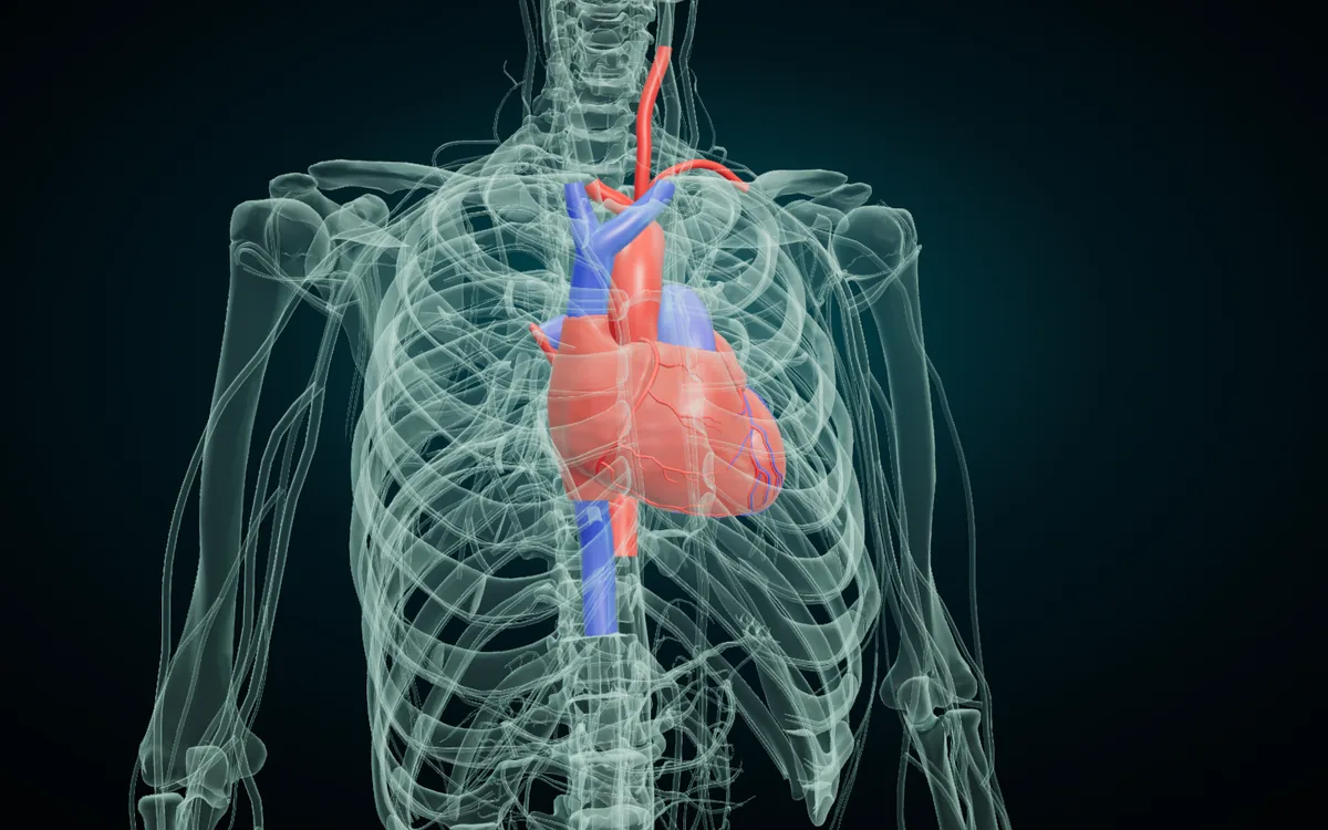
In this render the skeleton has turned to glass and the heart with its great vessels stays solid. The heart sits behind the lower sternum, and its apex points down and to the left. The superior vena cava drops in from above on the right, and the aorta arches up and back.
How blood flows through the heart
The heart is two pumps side by side. The right side sends blood to the lungs; the left side sends it to the rest of the body. The NHLBI describes the path in this order:
- Oxygen-poor blood from the body enters the right atrium through the superior and inferior venae cavae.
- It passes the tricuspid valve into the right ventricle.
- The right ventricle pumps it through the pulmonary valve into the pulmonary trunk and pulmonary arteries, to the lungs.
- Oxygen-rich blood comes back through the four pulmonary veins into the left atrium.
- It passes the mitral valve into the left ventricle.
- The left ventricle pumps it through the aortic valve into the aorta and out to the body.
venae cavae
tricuspid valve
pulmonary valve
pulmonary arteries
pulmonary veins
mitral valve
aortic valve
aorta
Read each row left to right. The top row is the pulmonary circuit, the bottom row the systemic circuit. The valves keep blood moving one way and stop it flowing back.
The atlas colours each vessel by the blood it carries. The pulmonary trunk and pulmonary arteries are blue, because they carry oxygen-poor blood to the lungs. The pulmonary veins are red, because they bring oxygen-rich blood back. Expect this exception in exams; the colours let you check it at a glance.
Chambers of the heart
| English | Latin | Arabic |
|---|---|---|
| Right atrium | Atrium dextrum | الأذين الأيمن |
| Right ventricle | Ventriculus dexter | البطين الأيمن |
| Left atrium | Atrium sinistrum | الأذين الأيسر |
| Left ventricle | Ventriculus sinister | البطين الأيسر |
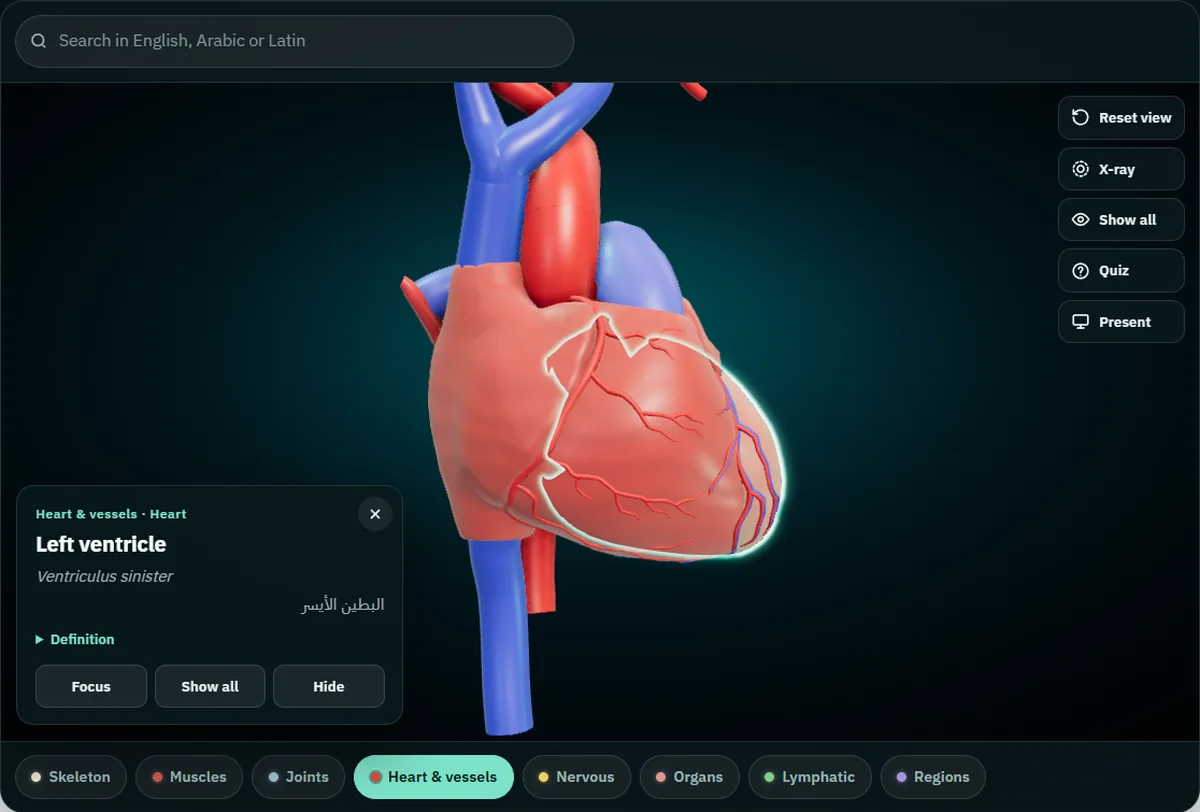
Tap the left ventricle and the card reads "Left ventricle", with Ventriculus sinister and البطين الأيسر under it, and the line "Heart & vessels · Heart" above. Atrium is Latin for "entrance hall", and the atria are where blood enters the heart. Ventriculus means "little belly".
From the front, most of what you see is the right ventricle. The right atrium forms the right border and the left ventricle most of the left border. Turn the model and the left atrium appears at the back: it forms the base of the heart, where the four pulmonary veins open into it (TeachMeAnatomy: surfaces and borders of the heart).
The left ventricle pumps blood through the whole body, while the right ventricle only pumps it through the lungs. The NHLBI notes that the left ventricle generates the force needed for the whole body, which is why its wall is the thickest of the four chambers.
Valves of the heart
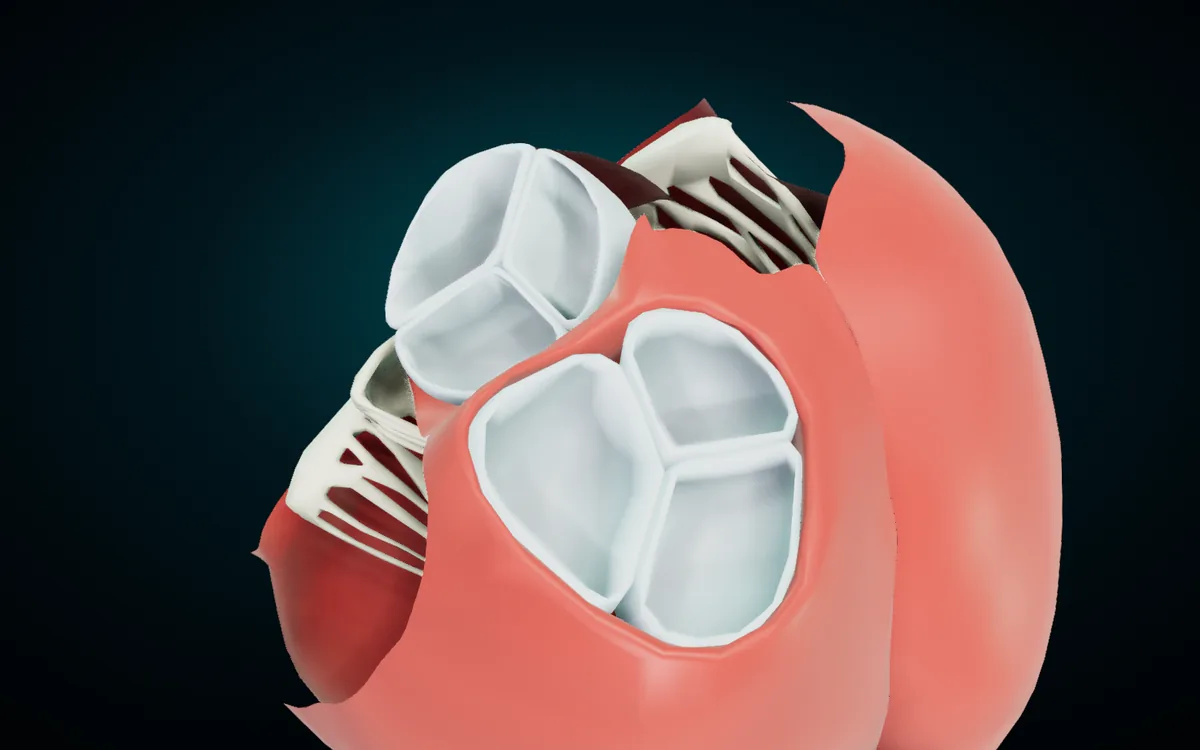
There are four valves in two pairs. The atrioventricular valves, tricuspid and mitral, sit between each atrium and its ventricle. The semilunar valves, pulmonary and aortic, sit where each ventricle opens into its artery (TeachMeAnatomy: heart valves).
| Valve | Latin | Arabic | Leaflets |
|---|---|---|---|
| Tricuspid valve | Valva atrioventricularis dextra | الصمام الأذيني البطيني الأيمن (ثلاثي الشرف) | 3 |
| Pulmonary valve | Valva trunci pulmonalis | الصمام الرئوي | 3 |
| Mitral valve | Valva atrioventricularis sinistra | الصمام الأذيني البطيني الأيسر (التاجي) | 2 |
| Aortic valve | Valva aortae | الصمام الأبهري | 3 |
The valve names in this table follow Terminologia Anatomica 2. The atlas models the valve leaflets, so a tap gives you a leaflet name. It has nine:
| English | Latin | Arabic |
|---|---|---|
| Septal leaflet of right atrioventricular valve | Cuspis septalis valvae atrioventricularis dextrae | الوريقة الحاجزية للصمام الأذيني البطيني الأيمن |
| Inferior leaflet of right atrioventricular valve | Cuspis inferior valvae atrioventricularis dextrae | الوريقة السفلية للصمام الأذيني البطيني الأيمن |
| Posterior leaflet of left atrioventricular valve | Cuspis posterior valvae atrioventricularis sinistrae | الوريقة الخلفية للصمام الأذيني البطيني الأيسر |
| Anterior semilunar leaflet of pulmonary valve | Valvula semilunaris anterior valvae trunci pulmonalis | الوريقة الهلالية الأمامية للصمام الرئوي |
| Right semilunar leaflet of pulmonary valve | Valvula semilunaris dextra valvae trunci pulmonalis | الوريقة الهلالية اليمنى للصمام الرئوي |
| Left semilunar leaflet of pulmonary valve | Valvula semilunaris sinistra valvae trunci pulmonalis | الوريقة الهلالية اليسرى للصمام الرئوي |
| Right coronary leaflet | Valvula coronaria dextra valvae aortae\* | الوريقة الإكليلية اليمنى |
| Left coronary leaflet | Valvula coronaria sinistra valvae aortae\* | الوريقة الإكليلية اليسرى |
| Non-coronary leaflet | Valvula noncoronaria valvae aortae | الوريقة غير الإكليلية |
Two leaflets are not modelled as separate parts: the anterior (superior) leaflet of the tricuspid valve and the anterior leaflet of the mitral valve. In the render above, the tricuspid and mitral openings carry only the leaflets the atlas models.
Your textbook may call the tricuspid's inferior leaflet the "posterior cusp". Both names are correct. Terminologia Anatomica 2 made "inferior leaflet" the official term and kept "posterior" as a synonym (FIPAT entry for this leaflet). The aortic leaflets work the same way. Their official names are right, left and posterior semilunar leaflets, and "right coronary", "left coronary" and "non-coronary" are official synonyms. The right and left coronary arteries start just above the first two, which is where those names come from.
A note for Arabic speakers: التاجي names the mitral valve, but many Arabic texts also call the coronary arteries الشرايين التاجية. The atlas uses الإكليلي for coronary, so الصمام التاجي is the mitral valve and الشريان الإكليلي is a coronary artery. Keep the two apart in an exam answer.
Papillary muscles
| English | Latin | Arabic |
|---|---|---|
| Anterior papillary muscle of right ventricle | Musculus papillaris anterior ventriculi dextri | العضلة الحليمية الأمامية للبطين الأيمن |
| Inferior papillary muscle of right ventricle | Musculus papillaris inferior ventriculi dextri | العضلة الحليمية السفلية للبطين الأيمن |
| Septal papillary muscle of right ventricle | Musculus papillaris septalis ventriculi dextri | العضلة الحليمية الحاجزية للبطين الأيمن |
| Inferior papillary muscle of left ventricle | Musculus papillaris inferior ventriculi sinistri | العضلة الحليمية السفلية للبطين الأيسر |
Papillary muscles rise from the ventricle walls and hold the atrioventricular leaflets through thin tendinous cords, the chordae tendineae. They contract while the ventricles contract, so the leaflets cannot flip back into the atria (TeachMeAnatomy). The right ventricle has three; the left ventricle has two, and the atlas models the inferior (posterior) one.
Great vessels
| English | Latin | Arabic |
|---|---|---|
| Superior vena cava | Vena cava superior | الوريد الأجوف العلوي |
| Inferior vena cava (thoracic part) | Vena cava inferior\* | الوريد الأجوف السفلي (الجزء الصدري) |
| Pulmonary trunk | Truncus pulmonalis | الجذع الرئوي |
| Right pulmonary artery | Arteria pulmonalis dextra | الشريان الرئوي الأيمن |
| Left pulmonary artery | Arteria pulmonalis sinistra | الشريان الرئوي الأيسر |
| Right superior pulmonary vein | Vena pulmonalis dextra superior | الوريد الرئوي العلوي الأيمن |
| Right inferior pulmonary vein | Vena pulmonalis dextra inferior | الوريد الرئوي السفلي الأيمن |
| Left superior pulmonary vein | Vena pulmonalis sinistra superior | الوريد الرئوي العلوي الأيسر |
| Left inferior pulmonary vein | Vena pulmonalis sinistra inferior | الوريد الرئوي السفلي الأيسر |
| Ascending aorta | Aorta ascendens | الأبهر الصاعد |
| Aortic arch | Arcus aortae | قوس الأبهر |
| Thoracic aorta | Aorta thoracica | الأبهر الصدري |
The two venae cavae bring blood from the body into the right atrium. The pulmonary trunk leaves the right ventricle and splits into the right and left pulmonary arteries; the atlas also has the split itself as a tappable part, "Bifurcation of pulmonary trunk". Four pulmonary veins, a superior and an inferior from each lung, end in the left atrium (TeachMeAnatomy: great vessels).
The aorta leaves the left ventricle as the ascending aorta, curves up and back as the arch, and runs down beside the spine as the thoracic aorta. The arch gives off three branches, which the atlas names too: the brachiocephalic trunk (Truncus brachiocephalicus, الجذع العضدي الرأسي), the left common carotid artery (الشريان السباتي الأصلي الأيسر) and the left subclavian artery (Arteria subclavia sinistra, الشريان تحت الترقوة الأيسر).
Coronary vessels: the heart's own blood supply
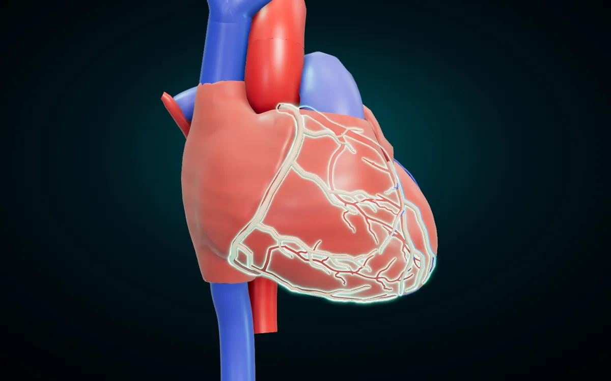
The coronary arteries branch off the aorta and carry oxygen-rich blood to the heart muscle itself. The left coronary artery feeds the left side of the heart through its circumflex and anterior descending branches; the right coronary artery feeds the right atrium and parts of both ventricles (NHLBI).
| English | Latin | Arabic |
|---|---|---|
| Right coronary artery | Arteria coronaria dextra | الشريان الإكليلي الأيمن |
| Right inferolateral branch of right coronary artery | Ramus inferolateralis dexter arteriae coronariae dextrae | الفرع السفلي الوحشي الأيمن للشريان الإكليلي الأيمن |
| Left coronary artery | Arteria coronaria sinistra | الشريان الإكليلي الأيسر |
| Anterior interventricular artery | Ramus interventricularis anterior\* | الشريان بين البطينين الأمامي |
| Septal branches of anterior interventricular artery | Rami septales interventriculares\* | الفروع الحاجزية للشريان بين البطينين الأمامي |
| Circumflex artery of heart | Arteria circumflexa cordis | الشريان المنعطف للقلب |
Clinicians call the anterior interventricular artery the LAD, short for left anterior descending artery. It runs down the groove between the two ventricles toward the apex, and its septal branches dive into the septum. The circumflex artery follows the groove between the atria and the ventricles, the coronary sulcus, around the left border to the back of the heart (TeachMeAnatomy: vasculature of the heart).
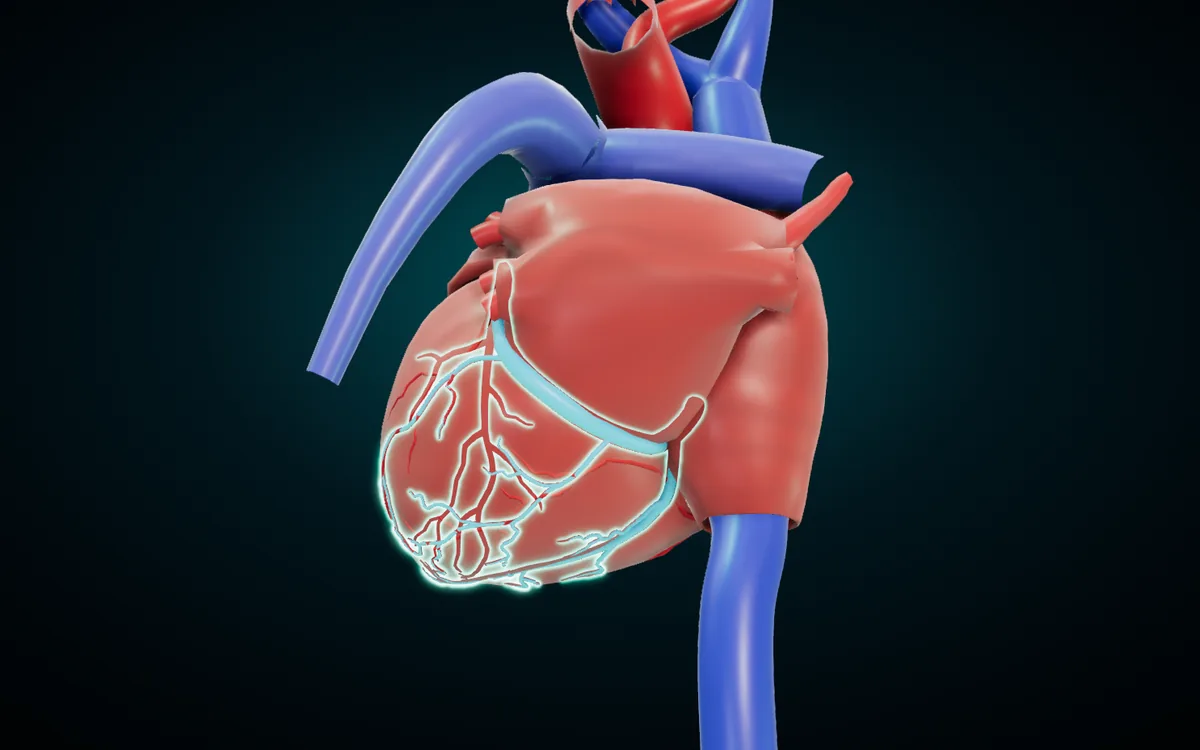
| English | Latin | Arabic |
|---|---|---|
| Great cardiac vein | Vena cardiaca magna\* | الوريد القلبي الكبير |
| Middle cardiac vein | Vena cardiaca media\* | الوريد القلبي الأوسط |
| Inferior vein of left ventricle | Vena ventriculi sinistri inferior\* | الوريد السفلي للبطين الأيسر |
| Coronary sinus | Sinus coronarius | الجيب الإكليلي |
The cardiac veins return oxygen-poor blood from the heart muscle. Most of them drain into the coronary sinus, the wide vein on the back of the heart, and the coronary sinus opens into the right atrium (FIPAT entry for the coronary sinus). The great cardiac vein climbs beside the anterior interventricular artery and the middle cardiac vein runs up the back, which is why Terminologia Anatomica also lists it as the posterior interventricular vein.
\* For these rows the Latin is the official form in Terminologia Anatomica 2. The atlas card currently shows an abbreviated or older spelling of the same term, or none for the inferior vena cava.
Wall and pericardium
| English | Latin | Arabic |
|---|---|---|
| Endocardium | Endocardium | شغاف القلب |
| Myocardium | Myocardium | عضل القلب |
| Epicardium | Epicardium | النخاب |
| Pericardium | Pericardium | التامور |
The heart wall has three layers. The endocardium lines the chambers and covers the valves; the myocardium is the thick layer of cardiac muscle that does the pumping; the epicardium covers the outside and is the inner (visceral) layer of the serous pericardium (TeachMeAnatomy: the heart wall). The pericardium is the sac around the heart. It holds the heart in place and a thin film of fluid between its layers reduces friction as the heart beats (NHLBI).
These layers and the pericardium are not separate parts in the atlas. The chambers are modelled as muscle shells, so you see the outside of each chamber but cannot peel the wall into layers. The conduction system (sinuatrial node, atrioventricular node and bundle branches) is not modelled either.
How to study the heart with the atlas
- Switch on Heart & vessels. Open the free 3D anatomy atlas, tap the Heart & vessels chip under the model and switch the other systems off. All eight systems are free in the browser with no account.
- Follow the blood. Tap the parts in the order of the six flow steps above: vena cava, right atrium, right ventricle, pulmonary trunk, pulmonary veins, left atrium, left ventricle, aorta. Saying each name out loud in English and Latin fixes the sequence.
- Search in the language you think in. The search box takes English, Latin or Arabic, so "left ventricle", "ventriculus sinister" and "البطين الأيسر" all find the same part.
- Open the heart. Hide the atria and the great arteries with the Hide button on each card, then look down at the base of the heart. That is how we made the valve render above.
- Use X-ray for position. Switch Skeleton on as well, select a chamber and press X-ray. The bones turn to glass and the part you selected stays lit, so you see where it sits behind the ribs.
- Quiz on what is on screen. Quiz asks ten "where is…?" questions drawn from the parts you can see. With only Heart & vessels on, the questions cover the heart and the vessels of the whole body; hide the parts you have not studied yet and the quiz skips them.
The same atlas covers the bones the heart sits behind and the muscles of the chest wall: see our guides to the bones of the human body and the muscles of the human body. If you teach, teaching anatomy with a projector shows Present mode, which puts the three names in large type on a classroom screen.
Frequently asked questions
How many chambers and valves does the heart have?
Four of each. The chambers are the right and left atria and the right and left ventricles. The valves are the tricuspid and mitral valves between the atria and ventricles, and the pulmonary and aortic valves at the exits of the ventricles.
What is the Latin name of the heart?
Cor. Its genitive cordis appears in terms such as Arteria circumflexa cordis. The adjective cardiacus, from the Greek kardia, appears in Vena cardiaca magna. The Arabic is القلب.
What is the difference between the tricuspid and the mitral valve?
The tricuspid valve sits between the right atrium and right ventricle and has three leaflets. The mitral valve sits between the left atrium and left ventricle and has two. Terminologia Anatomica calls them the right and left atrioventricular valves.
Why are the pulmonary arteries blue in the 3D model?
The atlas colours vessels by oxygen content. The pulmonary arteries carry oxygen-poor blood from the right ventricle to the lungs, so they are blue even though they are arteries. The pulmonary veins carry oxygen-rich blood back to the left atrium, so they are red.
What is the coronary sinus?
A short, wide vein in the groove on the back of the heart between the atria and the ventricles. It collects blood from most of the cardiac veins and empties into the right atrium.
Can I study the heart on my phone without internet?
Yes, with the Android app. Anatomy 3D on Google Play carries the same models and the same three-language names and works offline once installed. In the app the skeleton is free and the other systems have a free preview; the Anatomy 3D app page lists what it contains. The web atlas needs a connection.
Start with the four chambers
Learn the four chambers and the four valves first, then the vessels in the order blood meets them. Open the 3D heart in the free atlas, switch on Heart & vessels, and tap each part in the order of the flow list until you can name all three languages without the card.
3D renders: NasrTech, from Z-Anatomy models (CC BY-SA 4.0 licence).