

An adult skeleton is usually counted as 206 bones. This guide lists the main ones region by region, with three names for each: the English name from your textbook, the Latin term from Terminologia Anatomica, and the Arabic name used in Arabic medical teaching. The names in the tables match what the free 3D skeleton on NasrTech shows when you tap a bone, and every picture here is a render of that same model.
The 206 bones at a glance
| Region | What it contains | Bones |
|---|---|---|
| Skull | 8 cranial and 14 facial bones | 22 |
| Middle ear | malleus, incus and stapes on each side | 6 |
| Neck | hyoid bone | 1 |
| Vertebral column | 24 vertebrae, sacrum, coccyx | 26 |
| Thorax | sternum and 12 pairs of ribs | 25 |
| Shoulder girdles | clavicle and scapula on each side | 4 |
| Upper limbs | humerus, radius, ulna, 8 carpals, 5 metacarpals and 14 phalanges per side | 60 |
| Pelvic girdle | the two hip bones | 2 |
| Lower limbs | femur, patella, tibia, fibula, 7 tarsals, 5 metatarsals and 14 phalanges per side | 60 |
The first five rows form the axial skeleton: the 80 bones of the head, neck and trunk. The last four form the appendicular skeleton, the 126 bones of the girdles and limbs. The atlas uses the same split. Tap the femur and its card reads "Skeleton · Appendicular skeleton".
Skull bones: cranial and facial
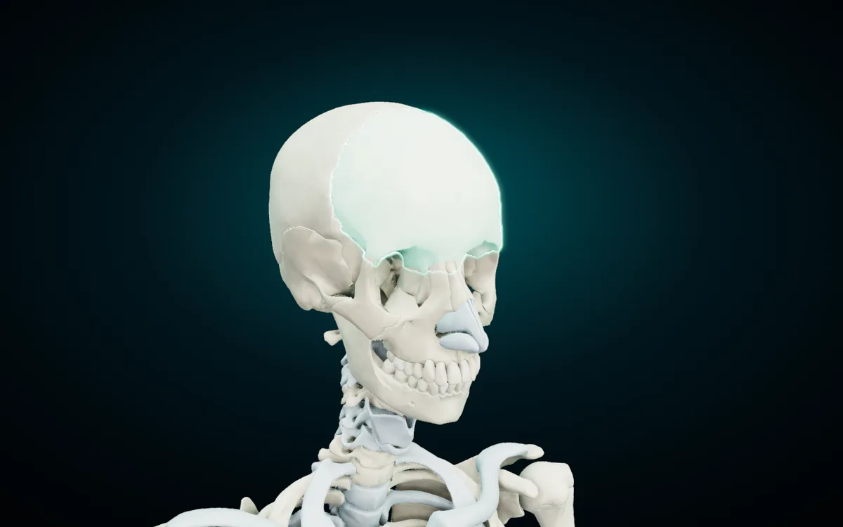
| English | Latin | Arabic |
|---|---|---|
| Frontal bone | Os frontale | العظم الجبهي |
| Parietal bone | Os parietale | العظم الجداري |
| Occipital bone | Os occipitale | العظم القذالي |
| Temporal bone | Os temporale | العظم الصدغي |
| Sphenoid bone | Os sphenoideum | العظم الوتدي |
| Ethmoid bone | Os ethmoideum | العظم الغربالي |
| Nasal bone | Os nasale | العظم الأنفي |
| Maxilla | Maxilla | الفك العلوي |
| Zygomatic bone | Os zygomaticum | العظم الوجني |
| Mandible | Mandibula | الفك السفلي |
| Hyoid bone | Os hyoideum | العظم اللامي |
The first six bones build the cranium, the case around the brain. The frontal, occipital, sphenoid and ethmoid bones are single and sit on the midline. The parietal and temporal bones come in pairs, and the atlas names each one with its side. The sphenoid and ethmoid are hard to picture from a flat drawing because most of each bone lies deep inside the skull. Hide the frontal bone and both parietal bones in the atlas, and you look down onto the floor of the cranium where those two sit.
The face takes 14 bones. The nasal bones, the maxillae and the zygomatic bones are paired. The mandible is single, and of the 22 skull bones it alone moves at a joint, the temporomandibular joint. The lacrimal bones, the palatine bones, the inferior nasal conchae and the vomer complete the facial count; search for them by name in the atlas. The hyoid sits in the neck below the mandible and touches no other bone. Muscles and ligaments hold it in place.
Spine, sternum and ribs
| English | Latin | Arabic |
|---|---|---|
| Manubrium of sternum | Manubrium sterni | قبضة القص |
| Body of sternum | Corpus sterni | جسم القص |
| Xiphoid process | Processus xiphoideus | الناتئ الرهابي |
| First rib | Costa prima | الضلع الأول |
| Sacrum | Os sacrum | العجز |
| Coccyx | Os coccygis | العصعص |
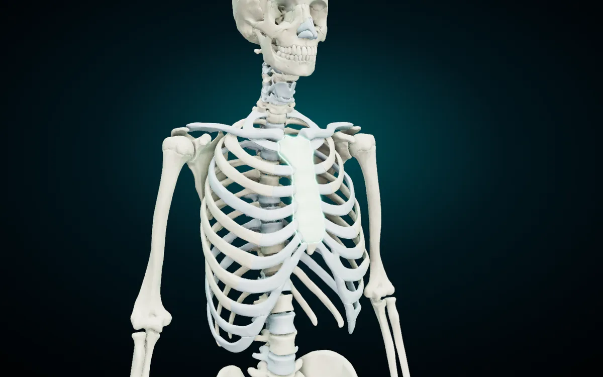
The sternum has three parts, and the atlas names each one: the manubrium at the top, where the clavicles and the first ribs attach; the body in the middle; and the small xiphoid process at the lower end. Behind it, 12 pairs of ribs curve back to the thoracic vertebrae. Ribs 1 to 7 reach the sternum through their own costal cartilage, so anatomists call them true ribs. Ribs 8 to 10 join the cartilage of the rib above (false ribs), and ribs 11 and 12 end free in the muscles of the flank (floating ribs).
The vertebral column has 7 cervical, 12 thoracic and 5 lumbar vertebrae, then the sacrum and the coccyx. The sacrum is five vertebrae fused into one bone, and the coccyx is usually four small ones fused together. That is how 33 vertebrae turn into 26 bones in the adult count. Two neck vertebrae carry names of their own: the atlas (C1) holds up the skull, and the axis (C2) gives it a pivot to turn on.
Upper limb bones, including the carpal bones
| English | Latin | Arabic |
|---|---|---|
| Clavicle | Clavicula | الترقوة |
| Scapula | Scapula | لوح الكتف |
| Humerus | Humerus | العضد |
| Radius | Radius | الكعبرة |
| Ulna | Ulna | الزند |
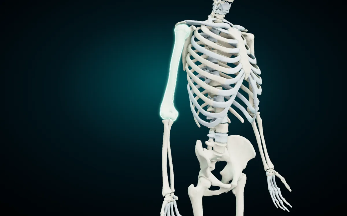
The clavicle and the scapula make the shoulder girdle. The humerus fills the arm, and the radius and ulna share the forearm. To keep the last two apart, put the radius on the thumb side and the ulna on the little-finger side. They mirror each other in shape: the radius is narrow at the elbow and wide at the wrist, the ulna wide at the elbow and narrow at the wrist. Isolate each one in the atlas and turn it, and the difference is plain.
The carpal bones
| English | Latin | Arabic |
|---|---|---|
| Scaphoid bone | Os scaphoideum | العظم الزورقي (الرسغ) |
| Lunate bone | Os lunatum | العظم الهلالي |
| Triquetrum bone | Os triquetrum | العظم المثلثي |
| Pisiform bone | Os pisiforme | العظم الحمصي |
| Trapezium bone | Os trapezium | العظم شبه المنحرف |
| Trapezoid bone | Os trapezoideum | العظم المنحرف |
| Capitate bone | Os capitatum | العظم الكبير |
| Hamate bone | Os hamatum | العظم الكلابي |
The wrist holds eight carpal bones in two rows. Starting from the thumb side, the proximal row runs scaphoid, lunate, triquetrum, pisiform, and the distal row runs trapezium, trapezoid, capitate, hamate. An English mnemonic keeps that order: "She Looks Too Pretty, Try To Catch Her".
Watch the trapezium and the trapezoid. The trapezium sits at the base of the thumb, and the trapezoid sits beside it toward the index finger. In Arabic one word separates them: العظم شبه المنحرف is the trapezium, العظم المنحرف the trapezoid.
Arabic has a second trap. It calls both the scaphoid of the wrist and the navicular of the foot العظم الزورقي, "the boat-shaped bone". The Greek skaphe and the Latin navicula both mean boat, so the two names say the same thing. The atlas adds (الرسغ), "wrist", to the scaphoid to keep them apart.
Past the carpus come five metacarpals and 14 phalanges: two in the thumb and three in each finger.
Pelvis and lower limb bones
| English | Latin | Arabic |
|---|---|---|
| Hip bone | Os coxae | عظم الورك |
| Femur | Os femoris | عظم الفخذ |
| Patella | Patella | الرضفة |
| Tibia | Tibia | الظنبوب |
| Fibula | Fibula | الشظية |
| Talus | Os tali | الكاحل |
| Calcaneus | Calcaneus | العقب |
| Navicular bone | Os naviculare | العظم الزورقي |
| Cuboid bone | Os cuboideum | العظم النردي |
| First metatarsal bone | Os primum metatarsi | عظم مشط القدم الأول |
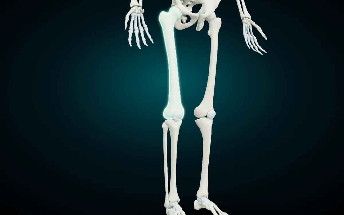
Each hip bone starts as three bones, the ilium, the ischium and the pubis, which fuse into one by adulthood. The two hip bones and the sacrum make the bony pelvis. The femur is the longest bone in the body. The patella is a sesamoid bone: it forms inside the tendon of the quadriceps and glides over the front of the femur. Below the knee, the tibia carries the weight on the inner side of the leg, and the slim fibula runs along the outer side.
The foot repeats the pattern of the hand with seven tarsal bones instead of eight. The talus sits on top and meets the tibia and fibula at the ankle joint. The calcaneus under it forms the heel. The navicular, the cuboid and three cuneiform bones lead on to five metatarsals and 14 phalanges, the same count as the hand.
Why anatomy uses three names for one bone
Latin is the international reference. Terminologia Anatomica gives one Latin term per structure, and English adjectives grow from it: Os femoris gives "femoral", Os coxae gives "coxal". English is the working language of most anatomy textbooks, and in many Arab countries medicine is taught in English, so students need the English and Arabic names side by side. The Arabic names in the atlas follow the conventions of the Unified Medical Dictionary (المعجم الطبي الموحد).
The three don't always line up word for word. Os coxae is "hip bone" in English and عظم الورك in Arabic, and older English books call it the innominate bone. The atlas gives the talus as الكاحل, while in everyday Arabic الكاحل means the whole ankle. A three-column table settles these gaps when your lecturer uses one name and your book uses another.
Paired bones also carry a side. In the English view of the atlas the card reads "Femur (right)"; in the Arabic view it reads "عظم الفخذ (يمين)". The Latin line shows the term without a side. If an exam asks for it in Latin, add dexter for right or sinister for left.
Learn them on a skeleton you can turn
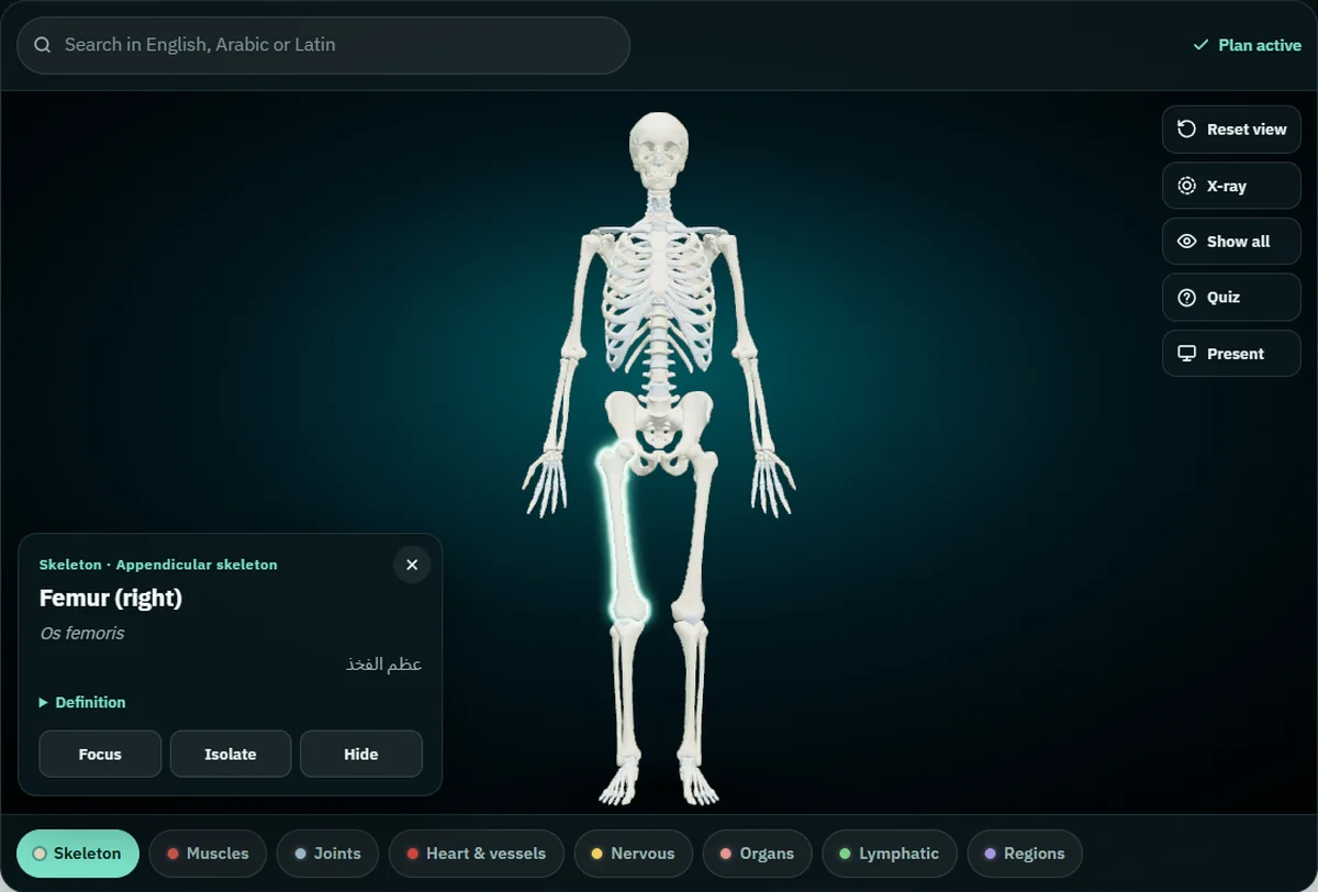
A table gives you the names. The model shows you where each bone sits and what touches it. The free 3D skeleton runs in Chrome, Edge, Safari or Firefox on a laptop, tablet or phone, and it needs no account. Five things to try with the tables above:
- Tap a bone. A card opens with the English name and side, the Latin term, the Arabic name, the group and a short definition in English.
- Double-tap to fly in. The camera moves close, which helps with small bones like the pisiform.
- Search in any of the three languages. Type femur, os femoris or عظم الفخذ and the model flies to the same bone.
- Isolate, hide or X-ray. Isolate shows one bone alone, Hide removes what is in the way, and X-ray turns the rest to glass.
- Run the quiz. Quiz asks ten "Where is the…?" questions, and you answer by tapping the model. It only asks about parts still on screen, so hide the bones you already know and let it test you on the rest.
Present mode fills the screen with the model and names each part you tap in large type in all three languages, which works for a study group around a TV. Lecturers can read how to use it in class in teaching anatomy with a projector. Once the bones feel solid, the names of the muscles of the human body follow the same three-column format; muscles sit in the paid part of the atlas.
Why the atlas shows 277 parts and textbooks say 206
Textbooks count 206 bones in an adult. The skeleton in the atlas lists 277 structures. Most of the difference comes from two choices. The atlas gives each tooth and each costal cartilage its own entry, though neither counts as a bone, and it splits the sternum into manubrium, body and xiphoid process. Dental students get the teeth on the same model as the jaws, and anyone else can hide them. When an exam asks how many bones the body has, the answer stays 206.
Quick answers
How many bones are in the human body? An adult has 206 by the usual count. A newborn has more separate pieces, because bones such as the hip bone and the sacrum form from several parts that fuse during growth.
Which is the largest bone, and which the smallest? The femur is the largest. The stapes, one of the three ossicles of the middle ear, is the smallest.
What are the axial and appendicular skeletons? The axial skeleton is the skull, hyoid, vertebral column and thorax. The appendicular skeleton is the shoulder and pelvic girdles plus the limbs.
Start with the free skeleton
Pick one region from this guide, open the 3D skeleton in your browser, and tap through its bones until you can name each one in the language your exam uses. Then hide the ones you know and run the quiz. If you study on your phone, Anatomy 3D for Android carries the same models and names, works offline, and adds cut planes on three axes; the Anatomy 3D app page has the details.
3D renders: NasrTech, from Z-Anatomy models (CC BY-SA 4.0).