

The arm has 4 muscles in two compartments. The forearm has 20 in two compartments and five layers. This guide lists every one of them where exam questions expect it: by compartment, then by layer, with the English name, the Latin term, the Arabic term, the main action and the nerve.
The English and Arabic names come from the cards of our free 3D Anatomy Atlas, which names each muscle, or each head of a muscle, when you tap it. Every picture here is a render of that model. Actions and nerves follow the TeachMeAnatomy muscle pages. If you want the 32 key muscles of the whole body first, start with our overview of the muscles of the human body and come back here for the upper limb.
The four compartments and their nerves
| Region | Compartment | Muscles | Nerve |
|---|---|---|---|
| Arm | Anterior (flexor) | Biceps brachii, coracobrachialis, brachialis | Musculocutaneous nerve |
| Arm | Posterior (extensor) | Triceps brachii | Radial nerve |
| Forearm | Anterior (flexor) | 8 muscles in 3 layers | Median nerve, except flexor carpi ulnaris and the medial half of flexor digitorum profundus (ulnar nerve) |
| Forearm | Posterior (extensor) | 12 muscles in 2 layers | Radial nerve and its deep branch (posterior interosseous nerve) |
Learn this table before any attachment: nerve-injury questions start from it. Three exceptions are worth knowing from day one: brachialis also gets a twig from the radial nerve, brachioradialis sits in the extensor compartment but bends the elbow, and the ulnar nerve takes one and a half muscles from the median nerve's territory in the front of the forearm.
A compartment is a group of muscles wrapped in deep fascia with its own nerve and its own job. In the arm, the deep fascia sends two sheets, the medial and lateral intermuscular septa, in to the humerus, and they split the arm into a front and a back. The front compartment flexes, the back one extends.
Arm: anterior compartment
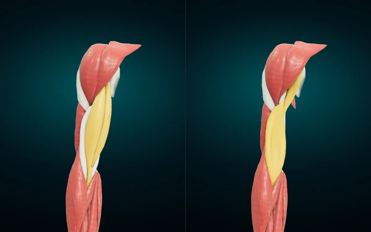
| English | Latin | Arabic | Main action | Nerve |
|---|---|---|---|---|
| Biceps brachii | Musculus biceps brachii | العضلة ذات الرأسين العضدية | Supinates the forearm, flexes the elbow, helps flex the shoulder | Musculocutaneous |
| Coracobrachialis | Musculus coracobrachialis | العضلة الغرابية العضدية | Flexes the arm at the shoulder, weak adductor | Musculocutaneous |
| Brachialis | Musculus brachialis | العضلة العضدية | Flexes the elbow | Musculocutaneous, with a contribution from the radial nerve |
The left half of the render is the arm as you see it from the front: biceps covers almost everything. On the right, biceps is hidden and the two deeper muscles appear. Coracobrachialis runs from the coracoid process of the scapula to the inner side of the humeral shaft, about halfway down. Brachialis starts on the shaft of the humerus and ends on the ulnar tuberosity, just below the elbow joint.
That attachment explains its action. Brachialis pulls on the ulna, and the ulna does not rotate, so brachialis bends the elbow in any position of the forearm. Biceps ends on the radial tuberosity of the radius instead. Pulling on the radius lets it rotate the forearm and turn the palm up as well as bend the elbow, which is why TeachMeAnatomy lists supination among its actions.
The atlas splits biceps into its two heads, each with its own card:
| Head | Latin | Arabic | Starts on |
|---|---|---|---|
| Long head of biceps brachii | Caput longum musculi bicipitis brachii | الرأس الطويل لذات الرأسين العضدية | Supraglenoid tubercle of the scapula |
| Short head of biceps brachii | Caput breve musculi bicipitis brachii | الرأس القصير لذات الرأسين العضدية | Coracoid process of the scapula |
Tap any of these in the atlas and the card's group line reads "Muscles · Anterior compartment of arm". The atlas uses the same compartment grouping you will see in your exam.
Arm: posterior compartment
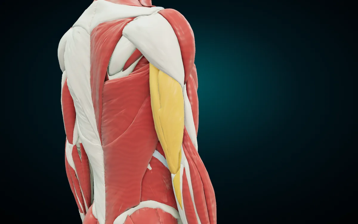
| English | Latin | Arabic | Main action | Nerve |
|---|---|---|---|---|
| Triceps brachii | Musculus triceps brachii | العضلة ثلاثية الرؤوس العضدية | Extends the elbow | Radial |
One muscle fills the whole back of the arm. Its three heads end on one tendon on the olecranon of the ulna, the point of your elbow.
| Head | Latin | Arabic | Starts on |
|---|---|---|---|
| Long head of triceps brachii | Caput longum musculi tricipitis brachii | الرأس الطويل لثلاثية الرؤوس العضدية | Infraglenoid tubercle of the scapula |
| Lateral head of triceps brachii | Caput laterale musculi tricipitis brachii | الرأس الوحشي لثلاثية الرؤوس العضدية | Humerus, above the radial groove |
| Medial head of triceps brachii | Caput mediale musculi tricipitis brachii | الرأس الإنسي لثلاثية الرؤوس العضدية | Humerus, below the radial groove |
The radial groove is the shallow spiral on the back of the humerus where the radial nerve runs. The lateral head starts above it and the medial head below it, so the nerve passes between the two heads. That is why a fracture of the shaft of the humerus is a classic radial nerve question. The long head is the only one that crosses the shoulder. TeachMeAnatomy notes that it is sometimes supplied by the axillary nerve instead.
Forearm: anterior compartment (flexors)
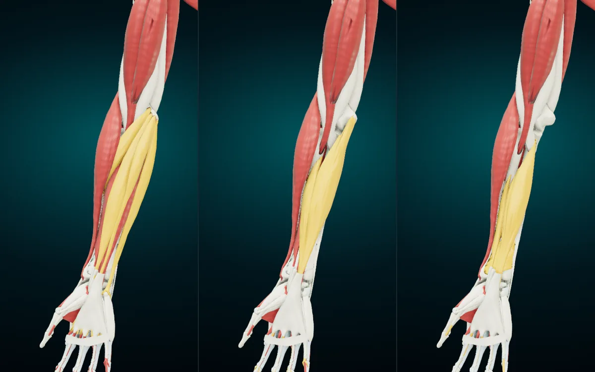
The front of the forearm holds 8 muscles in three layers. The render peels them in order: superficial, intermediate, deep. All the superficial muscles start from one common tendon on the medial epicondyle of the humerus, the bump on the inner side of your elbow (TeachMeAnatomy, anterior forearm).
Superficial layer
| English | Latin | Arabic | Main action | Nerve |
|---|---|---|---|---|
| Pronator teres | Musculus pronator teres | العضلة الكابة المدورة | Pronates the forearm | Median |
| Flexor carpi radialis | Musculus flexor carpi radialis | العضلة المثنية الكعبرية للرسغ | Flexes and abducts the wrist | Median |
| Palmaris longus | Musculus palmaris longus | العضلة الراحية الطويلة | Flexes the wrist | Median |
| Flexor carpi ulnaris | Musculus flexor carpi ulnaris | العضلة المثنية الزندية للرسغ | Flexes and adducts the wrist | Ulnar |
The table runs from the thumb side to the little-finger side, the same order you meet them on the forearm. The names carry the actions. "Carpi" means of the wrist, so a flexor carpi bends the wrist. "Radialis" and "ulnaris" give the side, and the side gives the second action: the radial flexor also tilts the hand toward the thumb (abduction) and the ulnar flexor tilts it toward the little finger (adduction). The Arabic follows the same logic: المثنية (flexor), الكعبرية (radial), للرسغ (of the wrist).
Palmaris longus is absent in about 15% of people. Flexor carpi ulnaris is the one superficial flexor the ulnar nerve supplies.
Intermediate layer
| English | Latin | Arabic | Main action | Nerve |
|---|---|---|---|---|
| Flexor digitorum superficialis | Musculus flexor digitorum superficialis | العضلة المثنية السطحية للأصابع | Flexes the four fingers at the knuckle and the middle joint; helps flex the wrist | Median |
One muscle forms this layer. It sends four tendons to the middle phalanges of the fingers, so it bends the proximal interphalangeal joints, the middle joints of the fingers. Its two heads, one from the humerus and ulna and one from the radius, leave a gap that the median nerve and the ulnar artery pass through.
Deep layer
| English | Latin | Arabic | Main action | Nerve |
|---|---|---|---|---|
| Flexor digitorum profundus | Musculus flexor digitorum profundus | العضلة المثنية العميقة للأصابع | The only muscle that flexes the fingertip joints; also flexes the knuckles and wrist | Medial half: ulnar. Lateral half: anterior interosseous branch of the median |
| Flexor pollicis longus | Musculus flexor pollicis longus | العضلة المثنية الطويلة للإبهام | Flexes the thumb at both of its joints | Anterior interosseous (median) |
| Pronator quadratus | Musculus pronator quadratus | العضلة الكابة المربعة | Pronates the forearm | Anterior interosseous (median) |
Superficialis and profundus are a pair worth learning together. Superficialis (السطحية) bends the middle joint of each finger, and profundus (العميقة) reaches past it to bend the last one. Try it on yourself: hold the middle joint of one finger straight with your other hand and bend only the tip. Only profundus can do that.
Pronator quadratus is the square muscle across the lower end of the radius and ulna. With pronator teres above it, the compartment has two pronators, one round (teres, المدورة) and one square (quadratus, المربعة).
Heads the atlas names separately
| Head in the atlas | Latin (Terminologia Anatomica) | Arabic |
|---|---|---|
| Superficial head of pronator teres | Caput humerale musculi pronatoris teretis | الرأس السطحي للكابة المدورة |
| Deep head of pronator teres | Caput ulnare musculi pronatoris teretis | الرأس العميق للكابة المدورة |
| Humeral head of flexor carpi ulnaris | Caput humerale musculi flexoris carpi ulnaris | الرأس العضدي للمثنية الزندية للرسغ |
| Ulnar head of flexor carpi ulnaris | Caput ulnare musculi flexoris carpi ulnaris | الرأس الزندي للمثنية الزندية للرسغ |
| Humero-ulnar head of flexor digitorum superficialis | Caput humero-ulnare musculi flexoris digitorum superficialis | الرأس العضدي الزندي للمثنية السطحية للأصابع |
| Radial head of flexor digitorum superficialis | Caput radiale musculi flexoris digitorum superficialis | الرأس الكعبري للمثنية السطحية للأصابع |
Textbooks call the two heads of pronator teres the humeral head and the ulnar head. The humeral head is the larger, more superficial one, so the atlas labels "superficial" and "deep" describe the same two heads.
A note on the Latin. The tables use the word order of Terminologia Anatomica, the international standard: flexor carpi radialis, flexor digitorum superficialis (IFAA entry). On some forearm cards the atlas currently puts the adjective before the noun it belongs to, as in flexor radialis carpi or flexor longus pollicis. Both orders name the same muscle; write the standard order in an exam.
Forearm: posterior compartment (extensors)
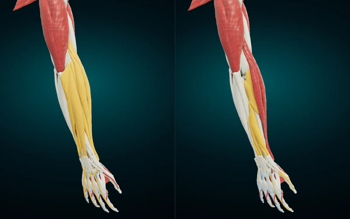
The back of the forearm holds 12 muscles in two layers. The radial nerve supplies all of them, most through its deep branch, which becomes the posterior interosseous nerve (TeachMeAnatomy, posterior forearm). In the atlas these muscles sit in the general "Muscles" group, so the card does not name the compartment for you. Use the table.
Superficial layer
| English | Latin | Arabic | Main action | Nerve |
|---|---|---|---|---|
| Brachioradialis | Musculus brachioradialis | العضلة العضدية الكعبرية | Flexes the elbow | Radial |
| Extensor carpi radialis longus | Musculus extensor carpi radialis longus | العضلة الباسطة الكعبرية الطويلة للرسغ | Extends and abducts the wrist | Radial |
| Extensor carpi radialis brevis | Musculus extensor carpi radialis brevis | العضلة الباسطة الكعبرية القصيرة للرسغ | Extends and abducts the wrist | Radial |
| Extensor digitorum | Musculus extensor digitorum | العضلة الباسطة للأصابع | Extends the four fingers | Deep branch of radial |
| Extensor digiti minimi | Musculus extensor digiti minimi | العضلة الباسطة للخنصر | Extends the little finger | Deep branch of radial |
| Extensor carpi ulnaris | Musculus extensor carpi ulnaris | العضلة الباسطة الزندية للرسغ | Extends and adducts the wrist | Deep branch of radial |
| Anconeus | Musculus anconeus | العضلة المرفقية | Helps extend the elbow and steadies it | Radial |
Brachioradialis is the odd one. It lives in the extensor compartment and has the extensors' nerve, yet its action is to bend the elbow. Expect it in "which muscle supplied by the radial nerve flexes the elbow?" questions.
The wrist extensors mirror the wrist flexors on the other side. Two radial extensors (longus and brevis) tilt the hand toward the thumb as they extend it, and one ulnar extensor tilts it toward the little finger. Extensor carpi ulnaris has a humeral and an ulnar head, and the atlas gives each its own card. Anconeus is small and triangular. It sits behind the elbow and continues the line of triceps. Its Arabic name المرفقية means "of the elbow".
Deep layer
| English | Latin | Arabic | Main action | Nerve |
|---|---|---|---|---|
| Supinator | Musculus supinator | العضلة الاستلقائية | Supinates the forearm | Deep branch of radial |
| Abductor pollicis longus | Musculus abductor pollicis longus | العضلة المبعدة الطويلة للإبهام | Abducts the thumb | Posterior interosseous |
| Extensor pollicis brevis | Musculus extensor pollicis brevis | العضلة الباسطة القصيرة للإبهام | Extends the thumb at its base joints | Posterior interosseous |
| Extensor pollicis longus | Musculus extensor pollicis longus | العضلة الباسطة الطويلة للإبهام | Extends all the joints of the thumb | Posterior interosseous |
| Extensor indicis | Musculus extensor indicis | العضلة الباسطة للسبابة | Extends the index finger | Posterior interosseous |
Four of the five deep muscles serve the thumb and the index finger. "Pollicis" means of the thumb (للإبهام) and "indicis" of the index finger (للسبابة). Supinator wraps around the upper radius and turns the palm up. In the right panel of the render the superficial extensors are hidden, and the deep muscles show as they run down the back of the forearm toward the thumb and the index finger.
Hand: the intrinsic muscles in brief
The muscles above start in the arm or forearm and send tendons into the hand. The hand also has small muscles that start and end inside it. The atlas includes them, and this table groups them by the TeachMeAnatomy hand page:
| Group | Muscles (English · Arabic) | Main action | Nerve |
|---|---|---|---|
| Thenar | Abductor pollicis brevis · العضلة المبعدة القصيرة للإبهام; flexor pollicis brevis · العضلة المثنية القصيرة للإبهام; opponens pollicis · العضلة المقابلة للإبهام | Move the thumb away from the palm, flex it and bring it across to touch the fingers | Median (the deep head of flexor pollicis brevis: ulnar) |
| Adductor pollicis | Oblique and transverse heads · العضلة المقربة للإبهام | Draws the thumb toward the palm | Ulnar |
| Hypothenar | Abductor digiti minimi, flexor digiti minimi, opponens digiti minimi · المبعدة والمثنية والمقابلة للخنصر | Move the little finger | Ulnar |
| Lumbricals | Lumbrical muscles of hand · العضلات الدودية لليد | Flex the knuckles and straighten the finger joints | Index and middle: median. Ring and little: ulnar |
| Interossei | Dorsal interossei · العضلات بين العظمية الظهرية لليد; palmar interossei · العضلات بين العظمية الراحية | Dorsal spread the fingers; palmar bring them together | Ulnar (deep branch) |
The short rule: the median nerve supplies the thenar muscles and the lumbricals of the index and middle fingers, and the ulnar nerve supplies the rest of the hand. The Latin names follow the forearm pattern: Musculus abductor pollicis brevis, Musculus opponens pollicis, Musculi lumbricales manus, Musculi interossei dorsales manus.
Find each muscle on the 3D model
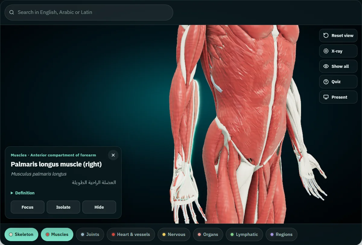
The screenshot shows the card you get when you tap palmaris longus: the English name and side, the Latin term, the Arabic term, and above them the system and group. Here is a routine that covers the whole upper limb in one sitting:
- Turn on Muscles. Open the atlas, keep Skeleton on and switch on Muscles in the bar under the model. Both are free in the browser with no account.
- Search in any of the three languages. "Pronator", "pronatoris" and "الكابة" all find pronator teres. The search box takes English, Latin or Arabic.
- Peel the layers in order. Tap a superficial flexor and press Hide on its card. Hide all four and flexor digitorum superficialis comes fully into view. Hide it and you reach the deep layer. The renders in this guide were made the same way.
- Isolate one muscle. Press Isolate on the card to see a muscle alone and turn it around. This is the fastest way to see where brachialis ends on the ulna.
- Quiz yourself. Quiz asks ten "where is…?" questions drawn from whatever is on screen, so hide what you have not studied yet and the questions stay on what remains.
Each arm muscle has a left and a right copy, and the card says which side you tapped. The bones these muscles move, the humerus, radius and ulna, have their own tables in our guide to the bones of the human body.
Frequently asked questions
How many muscles are in the forearm?
Twenty, by the grouping used here: 8 in the anterior compartment and 12 in the posterior compartment. Anconeus is counted with the superficial extensors, as TeachMeAnatomy does.
Which nerve supplies the muscles of the arm?
The musculocutaneous nerve supplies the anterior compartment (biceps brachii, coracobrachialis and brachialis). The radial nerve supplies the posterior compartment (triceps brachii). Brachialis also receives a small contribution from the radial nerve.
Which forearm flexors are not supplied by the median nerve?
Flexor carpi ulnaris and the medial half of flexor digitorum profundus. The ulnar nerve supplies both. Every other muscle in the anterior compartment of the forearm is supplied by the median nerve or its anterior interosseous branch.
Is brachioradialis a flexor or an extensor?
It flexes the elbow, but it belongs to the posterior (extensor) compartment of the forearm and the radial nerve supplies it.
What is the Arabic name of biceps and triceps?
Biceps brachii is العضلة ذات الرأسين العضدية, "the two-headed muscle of the arm". Triceps brachii is العضلة ثلاثية الرؤوس العضدية, "the three-headed muscle of the arm". Both Arabic names translate the Latin word for word.
Why does the atlas list heads instead of whole muscles?
Because exam questions often ask about one head: where the long head of triceps starts, or which head of flexor digitorum superficialis comes from the radius. The atlas gives each head its own English, Latin and Arabic name, so you can tap the exact part the question names.
Take it with you
The lower limb has its own guide to the pelvis and leg bones, with the same three-language tables.
The Anatomy 3D app for Android carries the same models and the same three-language names, works offline once installed, and draws each muscle's origin and insertion on the bone. If you teach this region, the guide to teaching anatomy with a projector shows Present mode, which puts the names in large type for a class.
Start with the compartment table, then open the free 3D atlas, hide the superficial flexors and see the deeper layers yourself.
3D renders: NasrTech, from Z-Anatomy models (CC BY-SA 4.0 licence). Actions and nerve supply: TeachMeAnatomy.