

There are 12 pairs of cranial nerves, numbered I to XII. They attach to the brain and brainstem rather than the spinal cord (XI is a partial exception, covered below), and they carry smell, sight, hearing, balance, taste, the feeling in the face, and the movements of the eyes, face, jaw, throat, tongue and neck. One of them, the vagus, also runs down into the chest and abdomen. This guide names all twelve in English, Latin and Arabic, says what each one does and where it leaves the skull, and then goes through the trigeminal branches and the brainstem nuclei that the free 3D Anatomy Atlas models. The names in the tables are the ones the atlas shows when you tap a nerve, and every picture here is a render of that same model.
The 12 cranial nerves at a glance
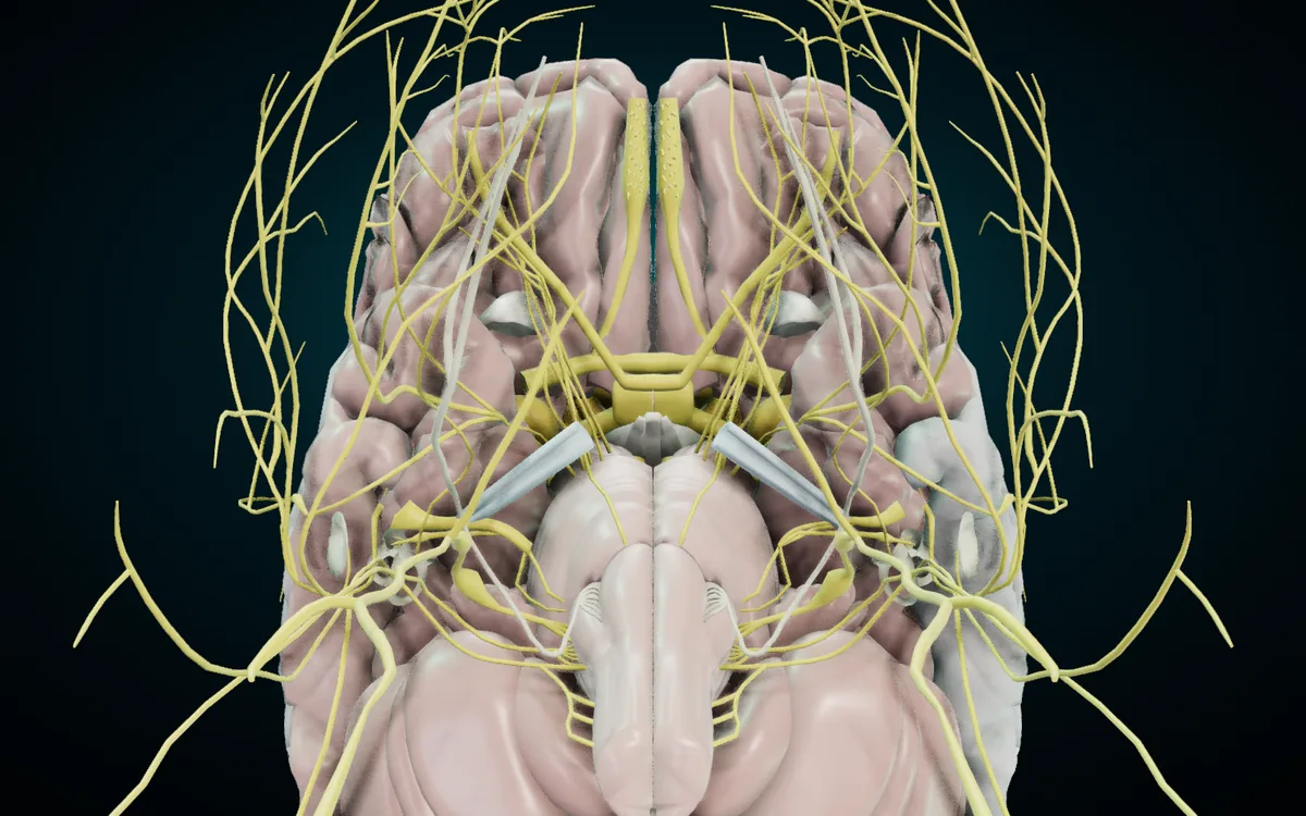
This is the brain seen from below, with the front at the top. The cranial nerves are yellow. The two olfactory nerves lie under the frontal lobes, the optic nerves meet at the optic chiasm in the centre, and below that the brainstem (pons and medulla) sends out the rest of the nerves on both sides, with the cerebellum at the bottom of the picture.
| No. | English | Latin | Arabic |
|---|---|---|---|
| I | Olfactory nerve | Nervus olfactorius | العصب الشمي |
| II | Optic nerve | Nervus opticus | العصب البصري |
| III | Oculomotor nerve | Nervus oculomotorius | العصب المحرك للعين |
| IV | Trochlear nerve | Nervus trochlearis | العصب البكري |
| V | Trigeminal nerve | Nervus trigeminus | العصب ثلاثي التوائم |
| VI | Abducens nerve | Nervus abducens | العصب المبعد |
| VII | Facial nerve | Nervus facialis | العصب الوجهي |
| VIII | Vestibulocochlear nerve | Nervus vestibulocochlearis | العصب الدهليزي القوقعي |
| IX | Glossopharyngeal nerve | Nervus glossopharyngeus | العصب اللساني البلعومي |
| X | Vagus nerve | Nervus vagus | العصب المبهم |
| XI | Accessory nerve | Nervus accessorius | العصب الإضافي |
| XII | Hypoglossal nerve | Nervus hypoglossus | العصب تحت اللسان |
The Latin terms match the Terminologia Anatomica (TA98) list of cranial nerves, which writes the number after each name in square brackets, for example Nervus vagus [X]. TA98 gives "abducent nerve" and "abducens nerve" as equal English names for VI. OpenStax and many other books call XI the "spinal accessory nerve", and an older name for VIII is the auditory nerve, which is the A in the rhyme further down.
Every cranial nerve is paired, so the atlas has a left and a right copy of each, and the card adds the side: "Vagus nerve (X) (right)".
What each cranial nerve does
| No. | Nerve | Type | Main job | Leaves the skull through |
|---|---|---|---|---|
| I | Olfactory | sensory | smell | cribriform plate of the ethmoid |
| II | Optic | sensory | vision | optic canal |
| III | Oculomotor | motor | four of the six eye muscles and the muscle that lifts the upper eyelid; constricts the pupil | superior orbital fissure |
| IV | Trochlear | motor | superior oblique muscle of the eye | superior orbital fissure |
| V | Trigeminal | both | feeling in the face, scalp, mouth and teeth; the muscles of mastication | V1 superior orbital fissure, V2 foramen rotundum, V3 foramen ovale |
| VI | Abducens | motor | lateral rectus muscle of the eye | superior orbital fissure |
| VII | Facial | both | muscles of facial expression; taste from the front two-thirds of the tongue; tear and salivary glands | internal acoustic meatus, then stylomastoid foramen |
| VIII | Vestibulocochlear | sensory | hearing and balance | internal acoustic meatus |
| IX | Glossopharyngeal | both | taste and feeling from the back third of the tongue; the stylopharyngeus muscle; the parotid gland | jugular foramen |
| X | Vagus | both | most muscles of the pharynx and larynx; the heart, lungs and most of the gut | jugular foramen |
| XI | Accessory | motor | sternocleidomastoid and trapezius | jugular foramen |
| XII | Hypoglossal | motor | the muscles of the tongue (all but palatoglossus) | hypoglossal canal |
The functions and exits follow the TeachMeAnatomy summary of the cranial nerves. The sensory, motor and mixed split is the one OpenStax Anatomy and Physiology uses: three nerves are only sensory (I, II, VIII), five are only motor (III, IV, VI, XI, XII), and four are mixed (V, VII, IX, X).
Two details come up in exams. First, the oculomotor nerve counts as motor but also carries parasympathetic fibres that make the pupil smaller. OpenStax names III, VII and IX as the nerves with autonomic fibres, and describes the vagus as almost entirely autonomic, so the parasympathetic set is III, VII, IX and X. Second, the tongue is shared out between four nerves: XII moves it, V3 (through the lingual nerve) gives general feeling to the front two-thirds, VII carries taste from the front two-thirds, and IX handles both taste and feeling at the back.
The nerves to the eye: II, III, IV and VI
The optic nerve carries vision from the retina. In the atlas it is one part, and the optic chiasm and optic tracts behind it are separate parts with their own cards (التصالب البصري and السبيل البصري in Arabic). You can see the chiasm, the X-shaped crossing, in the middle of the brain render above.
Three motor nerves share the six muscles that move each eye:
- Oculomotor nerve (III). The Radiopaedia article on the oculomotor nerve lists the superior, medial and inferior rectus, the inferior oblique and levator palpebrae superioris. Its parasympathetic fibres handle pupillary constriction and accommodation of the lens for near vision.
- Trochlear nerve (IV). The superior oblique only. The name comes from the trochlea, the small pulley its muscle's tendon runs through.
- Abducens nerve (VI). The lateral rectus only, which turns the eye outward, away from the nose. That outward turn is "abduction", hence the name, and the Arabic المبعد means the same thing.
The usual shorthand is LR6 SO4: lateral rectus by VI, superior oblique by IV, and III for the rest.
The trigeminal nerve (V) and its branches
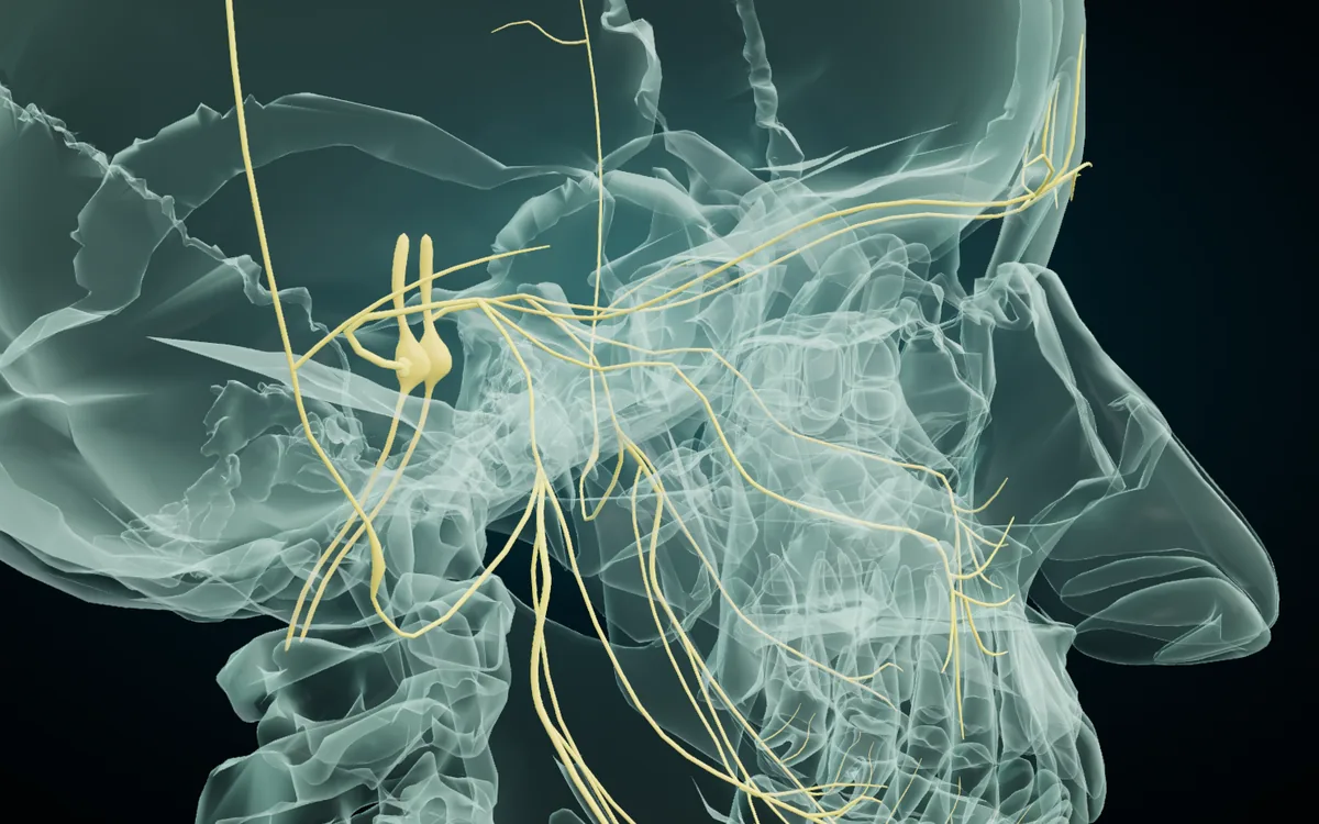
The trigeminal is the main sensory nerve of the face. Its name means "three twins", for its three divisions, and the Arabic ثلاثي التوائم keeps the same picture. The atlas models the nerve with its roots, all three divisions and several named branches of the mandibular division:
| English | Latin | Arabic |
|---|---|---|
| Trigeminal nerve (V) | Nervus trigeminus | العصب ثلاثي التوائم |
| Sensory root of trigeminal nerve | Radix sensoria nervi trigemini | الجذر الحسي للعصب ثلاثي التوائم |
| Motor root of trigeminal nerve | Radix motoria nervi trigemini | الجذر الحركي للعصب ثلاثي التوائم |
| Ophthalmic nerve (V1) | Nervus ophthalmicus | العصب العيني |
| Maxillary nerve (V2) | Nervus maxillaris | العصب الفكي العلوي |
| Meningeal branch of maxillary nerve | Ramus meningeus nervi maxillaris | الفرع السحائي للعصب الفكي العلوي |
| Anterior division of mandibular nerve | Divisio anterior nervi mandibularis | القسم الأمامي للعصب الفكي السفلي |
| Posterior division of mandibular nerve | Divisio posterior nervi mandibularis | القسم الخلفي للعصب الفكي السفلي |
| Buccal nerve | Nervus buccalis | العصب الخدي |
| Lingual nerve | Nervus lingualis | العصب اللساني |
| Inferior alveolar nerve | Nervus alveolaris inferior | العصب السنخي السفلي |
| Nerve to mylohyoid muscle | Nervus mylohyoideus | عصب العضلة الضرسية اللامية |
| Mental nerve | Nervus mentalis | العصب الذقني |
What the three divisions supply, from the TeachMeAnatomy cranial nerve summary:
- V1, ophthalmic. Sensation from the scalp, forehead and nose. Purely sensory.
- V2, maxillary. Sensation from the cheeks, lower eyelid, nasal mucosa, upper lip, upper teeth and palate. Purely sensory.
- V3, mandibular. Sensation from the front two-thirds of the tongue, the skin over the mandible and the lower teeth, plus motor fibres to the muscles of mastication. It is the only division with a motor job.
The mandibular branches in the table, as the TeachMeAnatomy page on the mandibular nerve describes them:
- Buccal nerve. Sensation from the lining of the cheek and the gums beside the lower molars and premolars.
- Inferior alveolar nerve. Sensation from the lower teeth. It leaves the mandible at the mental foramen as the mental nerve, which supplies the lower lip and chin.
- Nerve to mylohyoid. A branch of the inferior alveolar nerve with motor fibres to the mylohyoid and the anterior belly of the digastric.
- Lingual nerve. General sensation from the tongue. It also carries the taste and secretory fibres of the chorda tympani, a branch of the facial nerve.
Two notes on the Latin. TA98 lists the mandibular nerve as one nerve with its branches and has no separate "anterior division" or "posterior division" terms. The atlas uses them because its source model splits the nerve that way, and teaching sites such as Kenhub describe the same anterior and posterior divisions. TA98 also writes the two roots and the meningeal branch in short form, Radix motoria, Radix sensoria and Ramus meningeus, under their parent nerve. The atlas adds the parent nerve to the Latin so each term stands alone on its card.
Facial and vestibulocochlear nerves: VII and VIII
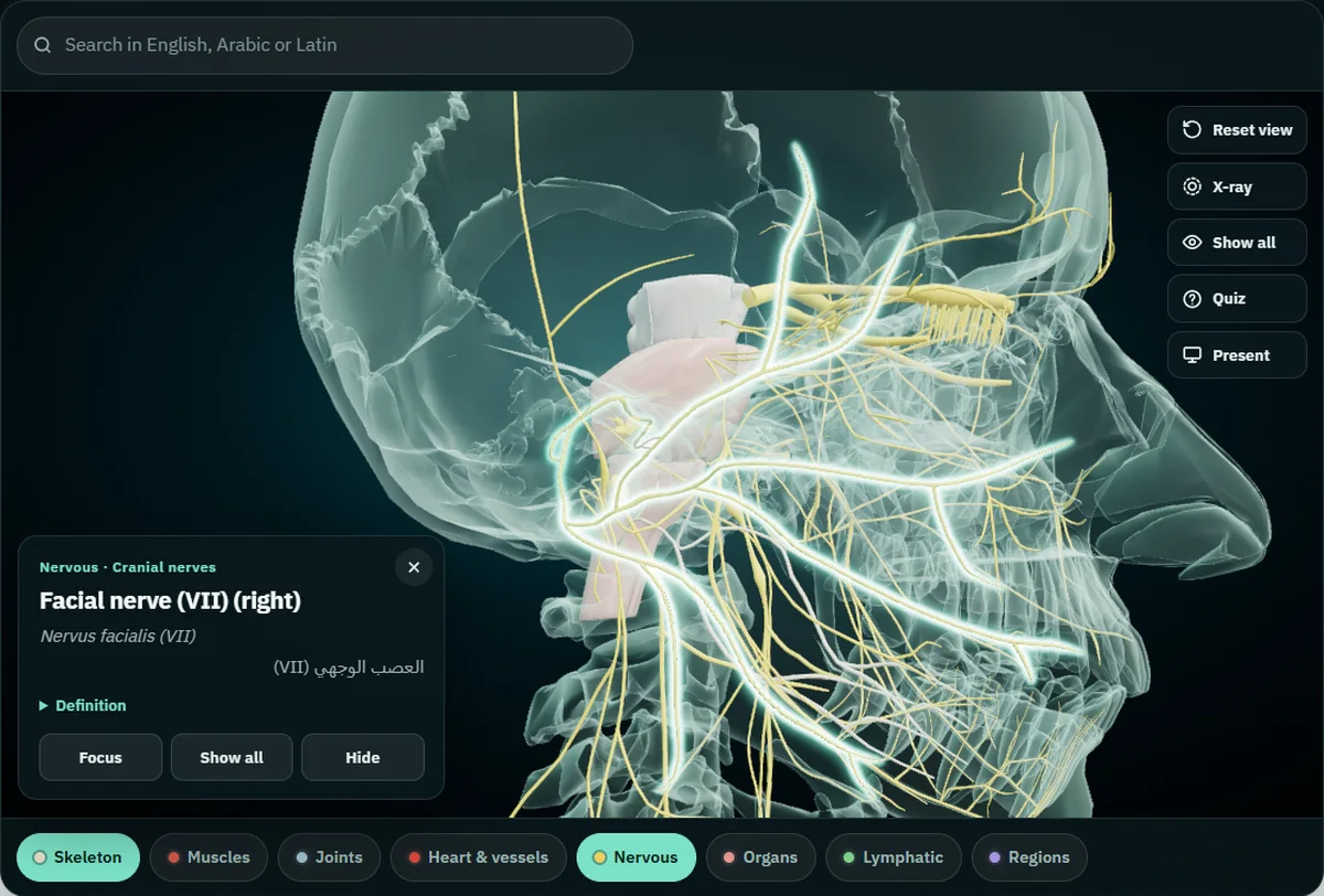
The facial nerve (VII) leaves the brainstem next to VIII, enters the internal acoustic meatus with it, and comes out of the skull at the stylomastoid foramen below the ear. From there it fans out over the face to the muscles of facial expression, which is what the highlighted nerve shows in the render. One of its branches has its own card in the atlas:
| English | Latin | Arabic |
|---|---|---|
| Chorda tympani | Chorda tympani | حبل الطبل |
The chorda tympani runs across the middle ear, which is where its name, "drum string", comes from, and then joins the lingual nerve. The TeachMeAnatomy facial nerve page describes it as carrying taste from the front two-thirds of the tongue, and the mandibular nerve page adds its parasympathetic fibres for the submandibular and sublingual salivary glands. In the face, the facial nerve ends inside the parotid gland by splitting into five branches: temporal, zygomatic, buccal, marginal mandibular and cervical.
The vestibulocochlear nerve (VIII) is two nerves running together, and the atlas lets you tap each one:
| English | Latin | Arabic | Carries |
|---|---|---|---|
| Vestibulocochlear nerve (VIII) | Nervus vestibulocochlearis | العصب الدهليزي القوقعي | hearing and balance |
| Cochlear nerve | Nervus cochlearis | العصب القوقعي | hearing, from the cochlea |
| Vestibular nerve | Nervus vestibularis | العصب الدهليزي | balance |
The cochlea, the vestibule, the tympanic membrane and the auditory tube are also in the atlas's Nervous system, so you can see where the cochlear and vestibular nerves are heading.
The last four: IX, X, XI and XII
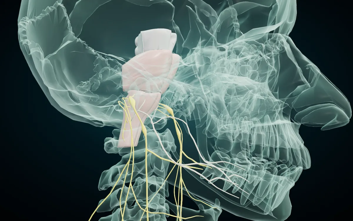
The glossopharyngeal (IX), vagus (X) and accessory (XI) nerves all leave the skull through the jugular foramen. The hypoglossal (XII) leaves through its own hypoglossal canal and curves forward under the jaw to the tongue. In the render the brainstem is the pale shape at the top left, and the four nerves come down from it into the neck.
- Glossopharyngeal (IX). Taste and sensation from the back third of the tongue, sensation from the middle ear, the carotid body and sinus, one muscle of the pharynx (stylopharyngeus) and the parotid gland.
- Vagus (X). Sensation and most of the muscles of the pharynx and larynx, and parasympathetic supply to the heart, lungs and most of the digestive tract.
- Accessory (XI). Two muscles of the neck and shoulder: sternocleidomastoid and trapezius. The Radiopaedia article on the accessory nerve lists the signs of damage to it: trouble shrugging the shoulder (trapezius) and turning the head (sternocleidomastoid).
- Hypoglossal (XII). All the muscles of the tongue except palatoglossus, which the vagus supplies through the pharyngeal plexus, per the Radiopaedia article on the hypoglossal nerve.
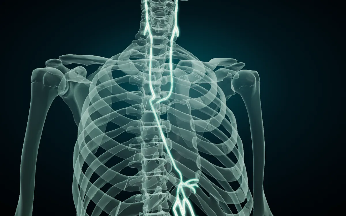
Vagus is Latin for "wandering", says the Radiopaedia article on the vagus nerve, because of its long path. The render shows why: isolate both vagus nerves with X-ray on and they run from the brainstem, down the neck and through the whole chest. The Arabic name المبهم means "vague", the English word that grew from the same Latin root. Radiopaedia also notes that the left and right vagus do not follow the same path in the thorax, and that near the oesophageal hiatus they form anterior and posterior trunks that pass into the abdomen.
Cranial nerve nuclei in the brainstem
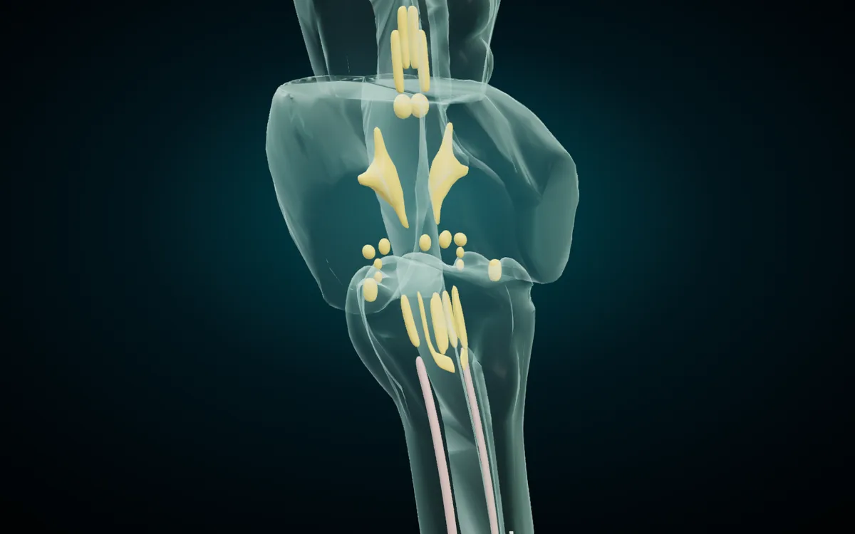
Cranial nerves III to XII have nuclei, groups of nerve cells where their motor fibres start or their sensory fibres end. Most sit in the brainstem. The atlas models 15 of these nuclei, each on both sides. Isolate them with X-ray on and the brainstem turns to glass around them, as above.
| English | Latin | Arabic | Nerve |
|---|---|---|---|
| Nucleus of oculomotor nerve | Nucleus nervi oculomotorii | نواة العصب المحرك للعين | III |
| Accessory nucleus of oculomotor nerve | Nucleus accessorius nervi oculomotorii | النواة الإضافية للعصب المحرك للعين | III (parasympathetic) |
| Nucleus of trochlear nerve | Nucleus nervi trochlearis | نواة العصب البكري | IV |
| Nucleus of abducens nerve | Nucleus nervi abducentis | نواة العصب المبعد | VI |
| Motor nucleus of facial nerve | Nucleus nervi facialis | النواة الحركية للعصب الوجهي | VII |
| Superior salivatory nucleus | Nucleus salivatorius superior | النواة اللعابية العلوية | VII |
| Anterior cochlear nucleus | Nucleus cochlearis anterior | النواة القوقعية الأمامية | VIII |
| Posterior cochlear nucleus | Nucleus cochlearis posterior | النواة القوقعية الخلفية | VIII |
| Vestibular nuclei | Nuclei vestibulares | النوى الدهليزية | VIII |
| Inferior salivatory nucleus | Nucleus salivatorius inferior | النواة اللعابية السفلية | IX |
| Nucleus of solitary tract | Nucleus tractus solitarii | نواة السبيل المنعزل | VII, IX, X |
| Nucleus ambiguus | Nucleus ambiguus | النواة الملتبسة | IX, X |
| Posterior nucleus of vagus nerve | Nucleus posterior nervi vagi | النواة الخلفية للعصب المبهم | X |
| Nucleus of accessory nerve | Nucleus nervi accessorii | نواة العصب الإضافي | XI |
| Nucleus of hypoglossal nerve | Nucleus nervi hypoglossi | نواة العصب تحت اللسان | XII |
The nerve column follows the nuclei listed for each nerve in the OpenStax cranial nerve table, with the vagal nuclei from Radiopaedia. The posterior nucleus of the vagus is the one many books call the dorsal motor nucleus of the vagus. The accessory nucleus of the oculomotor nerve is the parasympathetic part of III that the TA98 entry lists under the oculomotor nerve, and books usually call it the Edinger-Westphal nucleus. The nucleus of the accessory nerve sits in the upper spinal cord, not the brainstem: Radiopaedia places its cells in the ventral horn between C1 and C5, which is why the atlas files it under "Anterior horn of spinal cord".
What the atlas does not model
So you know what to look for in a textbook instead:
- The trigeminal nuclei. OpenStax places them in the midbrain, pons and medulla. The atlas has no separate parts for them.
- The ganglia. The trigeminal, geniculate, ciliary, pterygopalatine, otic and submandibular ganglia have no entries of their own.
- Most named branches. The trigeminal nerve and the chorda tympani are the exceptions. The five branches of the facial nerve in the face (temporal, zygomatic, buccal, marginal mandibular and cervical), the greater petrosal nerve, the recurrent laryngeal nerve and the auriculotemporal nerve have no entries of their own, so look them up by their parent nerve.
- The olfactory bulb and tract. There are no entries with these names; search for the olfactory nerve instead.
How to memorise the cranial nerves
Names in order. OpenStax gives the classic rhyme "On Old Olympus' Towering Tops, A Finn And German Viewed Some Hops": Olfactory, Optic, Oculomotor, Trochlear, Trigeminal, Abducens, Facial, Auditory (the old name for vestibulocochlear), Glossopharyngeal, Vagus, Spinal accessory, Hypoglossal.
Sensory or motor. A second rhyme gives the type in the same order, S for sensory, M for motor, B for both: "Some Say Marry Money But My Brother Says Brains Beauty Matter More". Check it against the table: I S, II S, III M, IV M, V B, VI M, VII B, VIII S, IX B, X B, XI M, XII M.
Let the Arabic explain the Latin. Several Arabic names translate the Latin meaning, so knowing one language helps with the other:
| Latin | Meaning | Arabic | Arabic word means |
|---|---|---|---|
| trochlea (IV) | pulley | البكري | بكرة, pulley |
| trigeminus (V) | three twins | ثلاثي التوائم | three twins |
| abducens (VI) | leading away | المبعد | the one that moves away |
| vagus (X) | wandering | المبهم | vague, undefined |
| hypoglossus (XII) | under the tongue | تحت اللسان | under the tongue |
Learn on the model. Read a name from the first table, find the nerve on the brain, tap it, and let the card confirm it.
Find each cranial nerve in the 3D atlas
The free 3D atlas runs in the browser on a laptop, tablet or phone, with no account. The skeleton loads first, and the cranial nerves are in the Nervous system. A route through all twelve:
- Turn on Nervous. Tap the Nervous chip under the model. You can leave the skeleton on for context.
- Search in any language. Type vagus, nervus vagus or المبهم, pick the result, and the model flies to the nerve and opens its card with the English name and side, the Latin term, the Arabic name and a short definition.
- Turn the brain over. Drag to rotate until you see the underside of the brain, as in the first render. Most of the twelve leave the brain on that surface.
- Isolate and X-ray. Select a nerve, press Isolate, then X-ray. The nerve stays solid and everything else turns to glass, so you can follow a nerve like the vagus or the hypoglossal through the skull and neck.
- Hide what is in the way. Hide the parts that cover a nerve, then press Show all to bring them back.
- Quiz yourself. Quiz asks ten "Where is the…?" questions and you answer by tapping the model. It draws from every part on screen, so isolate or hide down to the head first if you want the questions to stay on the cranial nerves.
Present mode shows each tapped name in large type in all three languages, which works for a study group around one screen or a lecturer at the front; the guide to teaching anatomy with a projector covers that setup. The Arabic version of the atlas has the same models with the interface in Arabic.
The cranial nerves pass through holes in the skull, so they are easier to learn once you know the bones. The guide to the skull bones in English, Latin and Arabic covers the cribriform plate, the sphenoid and the temporal bone. The muscles they supply, from the face to the tongue and neck, are in the muscles of the human body in English, Latin and Arabic, and the vagus nerve's main target in the chest is in the guide to the parts of the heart.
Frequently asked questions
How many cranial nerves are there?
Twelve pairs, numbered I to XII with Roman numerals. Each one has a left and a right nerve.
Which cranial nerves are only sensory?
Three: the olfactory (I), the optic (II) and the vestibulocochlear (VIII). Five are only motor (III, IV, VI, XI and XII), and four are mixed (V, VII, IX and X).
Which cranial nerves carry parasympathetic fibres?
The oculomotor (III), facial (VII), glossopharyngeal (IX) and vagus (X). III constricts the pupil, VII and IX drive the salivary glands, VII also drives the lacrimal gland, and X supplies the organs of the chest and abdomen.
Which cranial nerve is the longest?
The vagus (X). It is the one cranial nerve that reaches the organs of the chest and upper abdomen: it leaves the skull at the jugular foramen and runs down the neck, through the chest and into the abdomen. Its name is Latin for "wandering".
What is the Latin name of the facial nerve?
Nervus facialis. In Terminologia Anatomica it is written Nervus facialis [VII]. The Arabic name is العصب الوجهي.
Is the accessory nerve a cranial nerve if it starts in the spinal cord?
Yes. It is counted as cranial nerve XI. According to Radiopaedia, its fibres start in the upper spinal cord (C1 to C5), rise into the skull through the foramen magnum and leave again through the jugular foramen next to the vagus. It supplies sternocleidomastoid and trapezius.
Start with the base of the brain
Open the 3D brain and cranial nerves in your browser, turn on Nervous, and look at the brain from below. Start at the front with the olfactory and optic nerves, then work back through the brainstem to XII, saying the English, Latin and Arabic names as you tap each one. If you study on your phone, Anatomy 3D for Android carries the same models and names, works offline and adds cut planes on three axes; the Anatomy 3D app page has the details.
3D renders: NasrTech, from Z-Anatomy models (CC BY-SA 4.0).