

The tables below give 32 muscles you meet in the first weeks of anatomy, each with its English name, its Latin term and its Arabic term. We copied every name from the catalog of our 3D Anatomy Atlas, so when you tap a muscle there, the card shows the same words you read here. The Latin follows Terminologia Anatomica, and the Arabic follows the conventions of the Unified Medical Dictionary.
If you study in one language and sit exams in another, learn the three names together from the first week. The list suits students of medicine, dentistry, nursing, physiotherapy and sports science.
Twelve big muscles to learn first
| English | Latin | Arabic | Region |
|---|---|---|---|
| Deltoid | Musculus deltoideus | العضلة الدالية | Shoulder |
| Trapezius | Musculus trapezius | العضلة شبه المنحرفة | Neck and upper back |
| Pectoralis major | Musculus pectoralis major | العضلة الصدرية الكبيرة | Chest |
| Biceps brachii | Musculus biceps brachii | العضلة ذات الرأسين العضدية | Front of the arm |
| Triceps brachii | Musculus triceps brachii | العضلة ثلاثية الرؤوس العضدية | Back of the arm |
| Latissimus dorsi muscle | Musculus latissimus dorsi | العضلة الظهرية العريضة | Middle and lower back |
| Rectus abdominis muscle | Musculus rectus abdominis | العضلة المستقيمة البطنية | Abdominal wall |
| Diaphragm | Diaphragma | الحجاب الحاجز | Between chest and abdomen |
| Gluteus maximus muscle | Musculus gluteus maximus | العضلة الألوية الكبرى | Buttock |
| Rectus femoris muscle | Musculus rectus femoris | العضلة المستقيمة الفخذية | Front of the thigh |
| Biceps femoris | Musculus biceps femoris | العضلة ذات الرأسين الفخذية | Back of the thigh |
| Gastrocnemius | Musculus gastrocnemius | عضلة الساق (بطة الساق) | Calf |
These twelve draw the outline of the body: the cap of the shoulder, the chest, the front and back of the arm, the broad back, the belly wall, the breathing muscle, the buttock, the thigh and the calf. Seven of them appear in the atlas as separate heads or parts, each with its own three names. The atlas also keeps a left and a right copy of each paired muscle, and the card tells you which side you tapped.
Head and neck
| English | Latin | Arabic |
|---|---|---|
| Temporalis muscle | Musculus temporalis | العضلة الصدغية |
| Masseter | Musculus masseter | العضلة الماضغة |
| Orbicularis oculi | Musculus orbicularis oculi | العضلة الدويرية العينية |
| Sternocleidomastoid muscle | Musculus sternocleidomastoideus | العضلة القصية الترقوية الخشائية |
Temporalis and masseter close the jaw. Put your fingers on your temple and on the angle of your jaw, clench your teeth, and you feel both of them harden. Dental students study both in detail. Orbicularis oculi rings the eye and closes the eyelids; the atlas splits it into an orbital part and a palpebral part.
Sternocleidomastoid spells out its own attachments: sternum, clavicle and the mastoid process of the temporal bone. The Arabic name does the same, word by word: القصية (sternal), الترقوية (clavicular), الخشائية (mastoid). In the atlas you find it in the group "Accessory nerve (XI)", the cranial nerve that supplies it. Some groups in the atlas follow the nerve and others follow the compartment, so the small line above each name doubles as a revision cue.
Shoulder and arm
| English | Latin | Arabic |
|---|---|---|
| Brachialis muscle | Musculus brachialis | العضلة العضدية |
| Brachioradialis muscle | Musculus brachioradialis | العضلة العضدية الكعبرية |
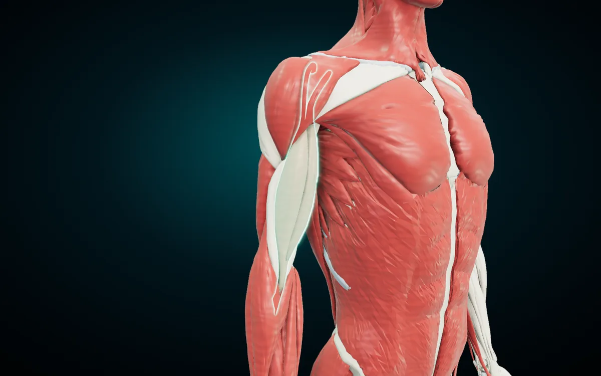
The render shows the three muscles that shape the arm from the front. The deltoid caps the shoulder; its name comes from the Greek letter delta, and the Arabic الدالية comes from the same letter, دال. Biceps brachii fills the front of the arm and bends the elbow. Triceps brachii fills the back and straightens it.
Brachialis lies under the biceps and is the main flexor of the elbow. Brachioradialis runs from the lower end of the humerus to the radius, which is what its name says: brachium (arm) plus radius. The atlas groups biceps and brachialis under "Anterior compartment of arm", the same label the name card shows when you tap either one.
Chest and abdomen
| English | Latin | Arabic |
|---|---|---|
| Pectoralis minor muscle | Musculus pectoralis minor | العضلة الصدرية الصغيرة |
| Serratus anterior muscle | Musculus serratus anterior | العضلة المنشارية الأمامية |
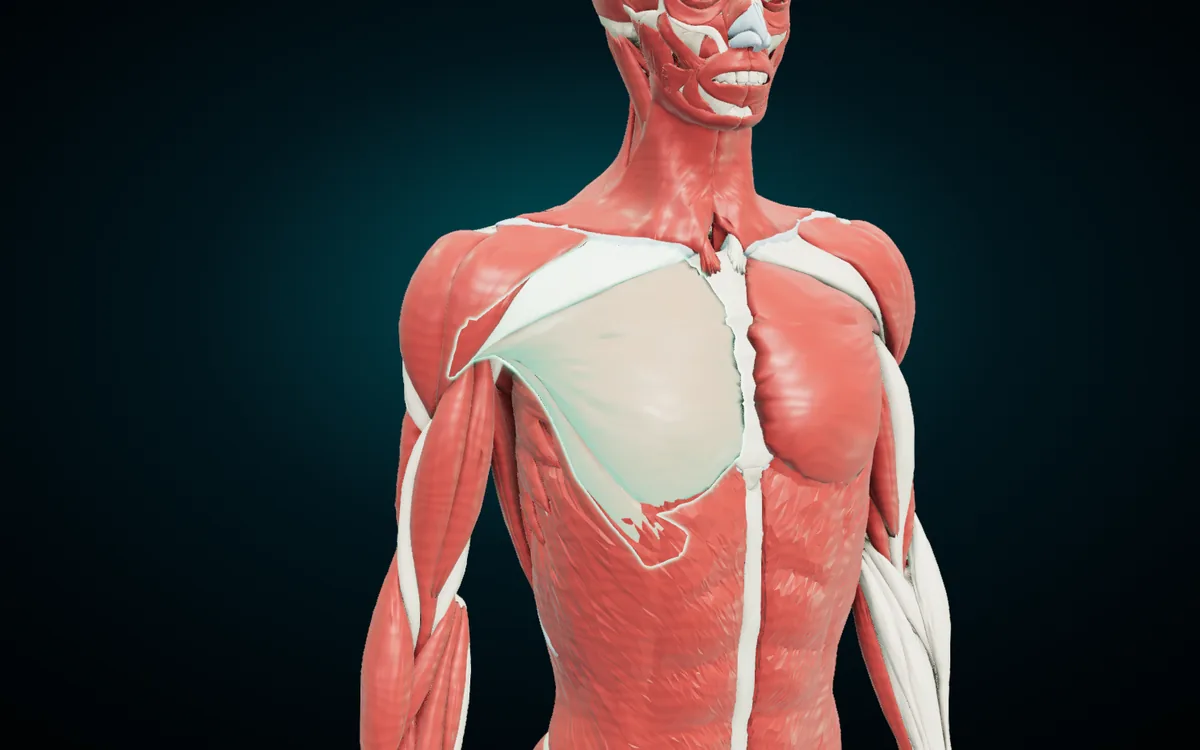
Pectoralis major covers the front of the chest and pulls the arm across the body. Pectoralis minor lies under it: tap the major, press Hide on the card, and the minor appears. Serratus anterior sits on the side of the rib cage in saw-tooth slips, which gives it its name in both languages (serratus, المنشارية). The atlas lists it under "Long thoracic nerve".
Rectus abdominis runs down the front of the abdomen on each side of the midline. The diaphragm closes the floor of the chest and is the main muscle of breathing. It is one of the few muscles the atlas lists once, with no left and right copy.
Back
| English | Latin | Arabic |
|---|---|---|
| Infraspinatus muscle | Musculus infraspinatus | العضلة تحت الشوكية |
| Teres major muscle | Musculus teres major | العضلة المدورة الكبيرة |
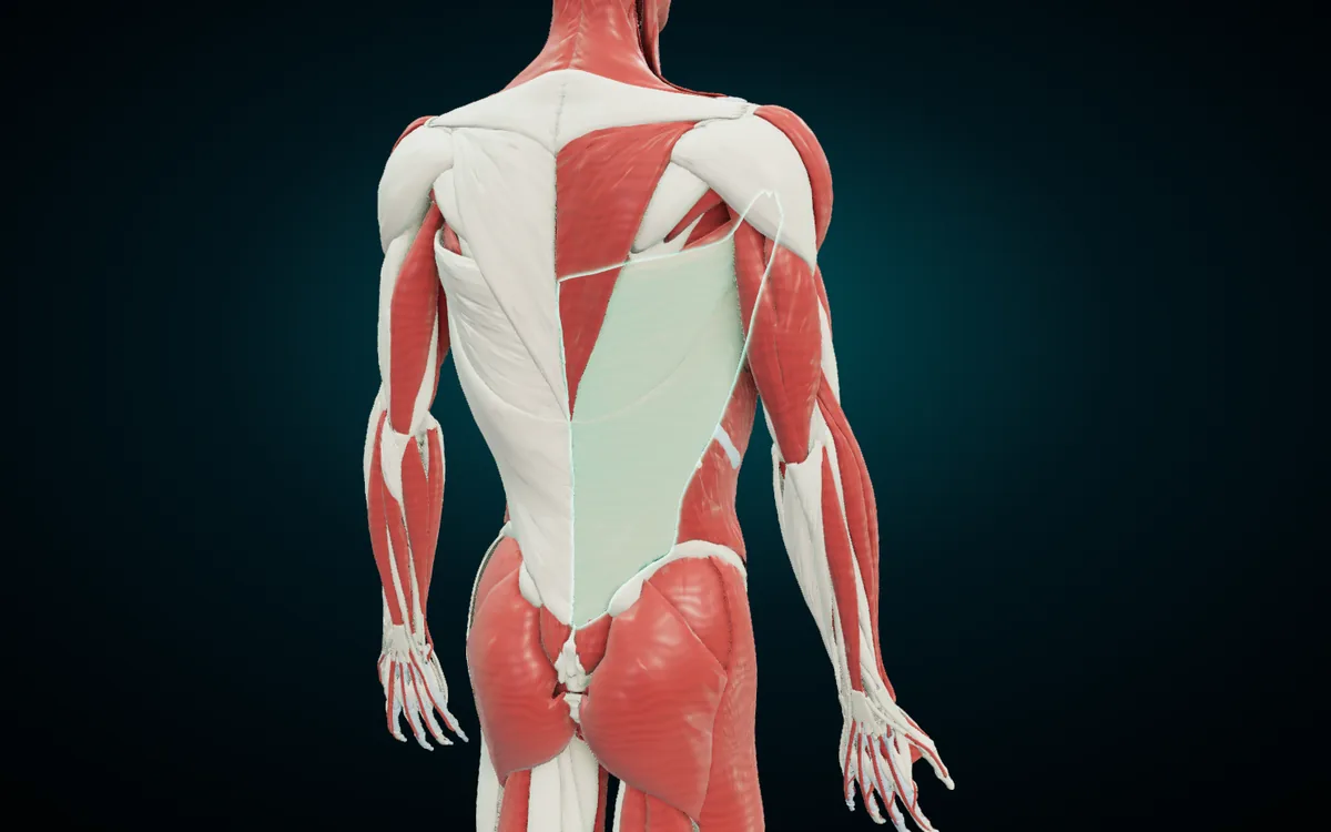
Trapezius covers the neck and upper back, and the atlas splits it into a descending, a transverse and an ascending part. Latissimus dorsi means "widest of the back", and العضلة الظهرية العريضة says the same thing in Arabic. It pulls the raised arm down and back, the movement of a pull-up.
Infraspinatus sits below the spine of the scapula: infra (below) plus spina. The Arabic تحت الشوكية is a word-for-word translation. Teres major, "large round", runs from the lower corner of the scapula to the humerus.
Hip and thigh
| English | Latin | Arabic |
|---|---|---|
| Gluteus medius muscle | Musculus gluteus medius | العضلة الألوية الوسطى |
| Iliacus | Musculus iliacus | العضلة الحرقفية |
| Psoas major | Musculus psoas major | العضلة القطنية الكبيرة |
| Sartorius muscle | Musculus sartorius | العضلة الخياطية |
| Vastus lateralis muscle | Musculus vastus lateralis | العضلة المتسعة الوحشية |
| Vastus medialis muscle | Musculus vastus medialis | العضلة المتسعة الإنسية |
| Adductor longus | Musculus adductor longus | العضلة المقربة الطويلة |
| Semitendinosus muscle | Musculus semitendinosus | العضلة نصف الوترية |
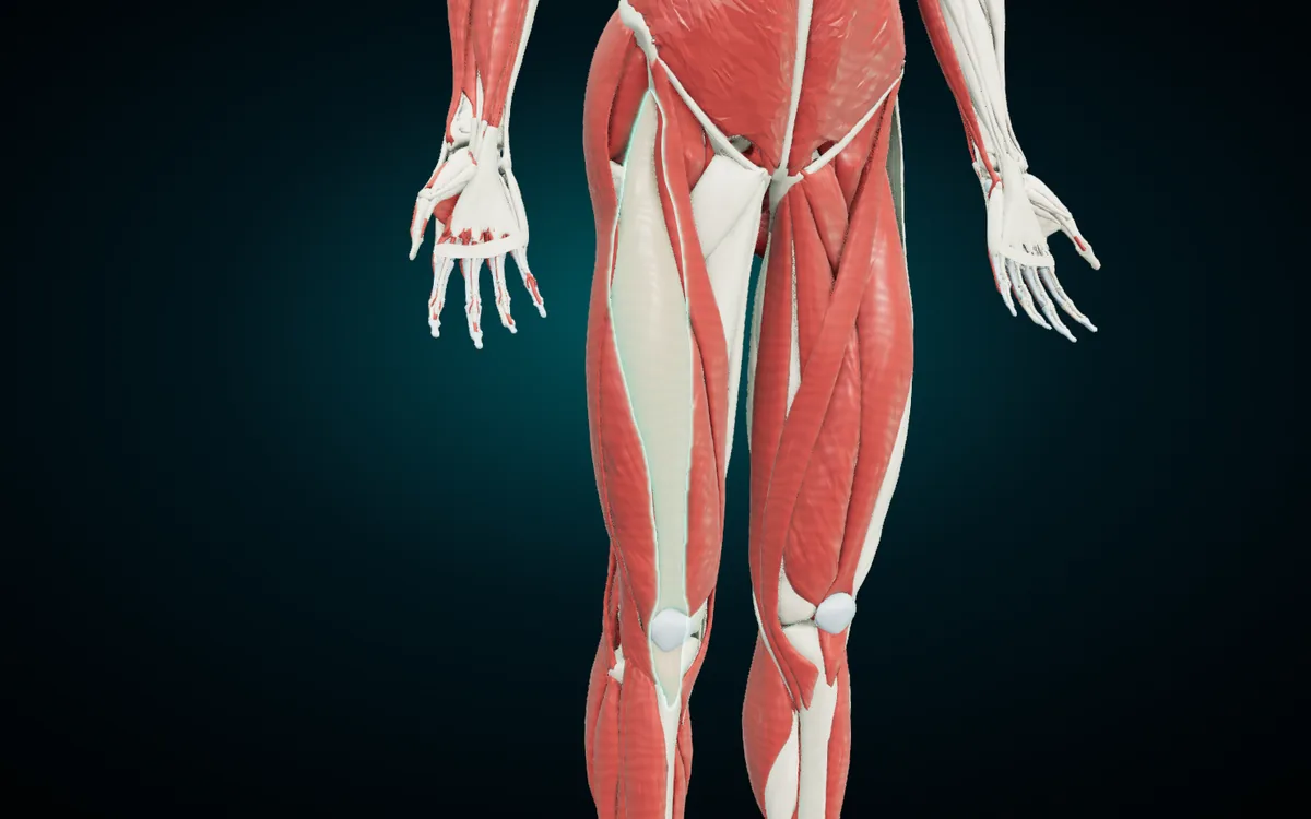
Rectus femoris is one of the four heads of the quadriceps. The other three are vastus lateralis, vastus medialis and vastus intermedius, which lies under rectus femoris. All four straighten the knee through the patella. Of the four, rectus femoris alone also crosses the hip, so it flexes the hip as well.
Sartorius crosses the front of the thigh on a diagonal, from the hip bone to the inner side of the knee. It is the longest muscle in the body, and both names mean "tailor's muscle". Iliacus and psoas major join to flex the hip. Gluteus maximus extends the hip, and gluteus medius keeps the pelvis level when you stand on one leg. At the back of the thigh, biceps femoris and semitendinosus belong to the hamstrings, which bend the knee.
Leg
| English | Latin | Arabic |
|---|---|---|
| Soleus muscle | Musculus soleus | العضلة النعلية |
| Tibialis anterior muscle | Musculus tibialis anterior | العضلة الظنبوبية الأمامية |
Gastrocnemius forms the bulge of the calf with its two heads, and soleus lies under it. Both end in the calcaneal tendon on the heel bone and point the foot down. Tibialis anterior runs along the front of the tibia and lifts the foot. The bones these muscles pull on, from the tibia to the calcaneus, have their own tables in our guide to the bones of the human body.
Why the atlas names heads and parts separately
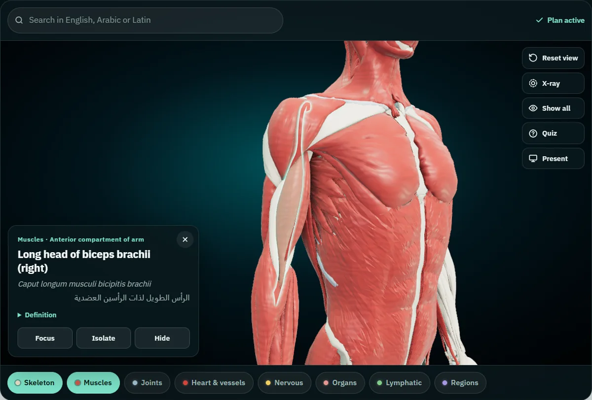
Tap the biceps in the atlas and the card reads "Long head of biceps brachii (right)", with Caput longum musculi bicipitis brachii and الرأس الطويل لذات الرأسين العضدية under it. Tap closer to the chest and you get the short head. The two heads start in different places: the long head above the shoulder socket, on the supraglenoid tubercle, and the short head on the coracoid process of the scapula. They join and end on the radius. "Biceps" means "two heads", and ذات الرأسين says the same.
| Part in the atlas | Latin | Arabic |
|---|---|---|
| Long head of biceps brachii | Caput longum musculi bicipitis brachii | الرأس الطويل لذات الرأسين العضدية |
| Short head of biceps brachii | Caput breve musculi bicipitis brachii | الرأس القصير لذات الرأسين العضدية |
| Clavicular part of deltoid muscle | Pars clavicularis musculi deltoidei | الجزء الترقوي من العضلة الدالية |
| Acromial part of deltoid muscle | Pars acromialis musculi deltoidei | الجزء الأخرمي من العضلة الدالية |
| Scapular spinal part of deltoid muscle | Pars spinalis scapularis musculi deltoidei | الجزء الشوكي الكتفي من العضلة الدالية |
| Clavicular head of pectoralis major muscle | Pars clavicularis musculi pectoralis majoris | الرأس الترقوي للعضلة الصدرية الكبيرة |
| Sternocostal head of pectoralis major muscle | Pars sternocostalis musculi pectoralis majoris | الرأس القصي الضلعي للعضلة الصدرية الكبيرة |
| Medial head of gastrocnemius | Caput mediale musculi gastrocnemii | الرأس الإنسي لعضلة الساق (بطة الساق) |
| Lateral head of gastrocnemius | Caput laterale musculi gastrocnemii | الرأس الوحشي لعضلة الساق (بطة الساق) |
The deltoid shows why the split matters. Its clavicular part raises the arm forward, the acromial part lifts it out to the side, and the scapular spinal part draws it back. When a question asks which fibres of the deltoid abduct the arm, the answer is a part, and the atlas lets you tap that exact part. The same logic covers the long, lateral and medial heads of triceps, the two heads of biceps femoris, the superficial and deep parts of masseter, and the three parts of trapezius.
The split also explains the atlas count. Its muscle system holds 683 structures, because left and right count separately, heads count one by one, and bursae and fasciae sit in the same system. Textbooks put the number of skeletal muscles at more than 600, and the total depends on how you count.
See a muscle against the bones with X-ray
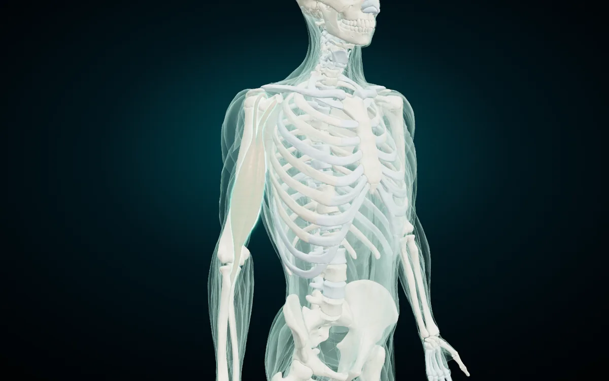
Select a muscle, then press X-ray. The other muscles turn to glass, the skeleton shows through, and your muscle stays lit. In the render the biceps spans both the shoulder and the elbow, which is why it acts on both joints. Try the same with rectus femoris and you see it cross the hip and the knee. Keep Skeleton and Muscles switched on together for this view.
How to study the muscles with the atlas
- Start with the bones, free. The whole skeleton, 277 structures including the teeth and the costal cartilages, opens in the browser with every tool and no account. Muscle names lean on bone names (sternocleidomastoid, brachioradialis, tibialis anterior), so the bones make the muscles easier.
- Unlock the muscles. The muscle system belongs to the paid plan with the six other systems: $4.99 a month or $24.99 a year, paid through Gumroad, which emails you a licence key. You can cancel any time.
- Search in the language you think in. The search box takes English, Latin or Arabic, so "biceps", "bicipitis" and "ذات الرأسين" all find the same muscle.
- Peel the layers. Hide a surface muscle to see the one beneath, isolate a muscle to turn it alone, and double-tap it to fly in.
- Quiz on what you show. Quiz asks ten "where is…?" questions drawn from whatever is on screen. Switch off every system except Muscles and hide the muscles you have not covered yet; the questions stay on what remains.
Study on your phone, offline
The Android app Anatomy 3D on Google Play carries the same models and the same three-language names, and it works without a connection once installed. For muscles it adds origins and insertions, drawn on the bone where each muscle starts and ends. It also has cut planes on three axes and can mirror to a TV. Purchases in the app are separate from the web plan, and the Anatomy 3D app page shows what the app contains.
Open the free 3D skeleton today and learn the bones of one region, then come back to its muscles. If you teach, the guide to teaching anatomy with a projector shows Present mode, which puts the three names in large type on a projector or TV.
3D renders: NasrTech, from Z-Anatomy models (CC BY-SA 4.0 licence).