

Each lower limb has 30 bones: the femur, the patella, the tibia, the fibula, 7 tarsals, 5 metatarsals and 14 phalanges. Add the hip bone that attaches it to the trunk and you have 31 per side, 62 for both. This guide names every one of them in English, Latin and Arabic, lists the landmarks that come up in exams, and shows each bone on a 3D model. The names in the tables are the ones the free 3D Anatomy Atlas shows when you tap a bone, and every picture is a render of that same model.
This page is part of a series. The bones of the human body in English, Latin and Arabic gives the whole skeleton in one table, and the guide to all 22 skull bones covers the head. Here the focus is the pelvis, the leg and the foot.
The bones of one lower limb at a glance
| English | Latin | Arabic | Per side |
|---|---|---|---|
| Hip bone | Os coxae | عظم الورك | 1 |
| Femur | Os femoris | عظم الفخذ | 1 |
| Patella | Patella | الرضفة | 1 |
| Tibia | Tibia | الظنبوب | 1 |
| Fibula | Fibula | الشظية | 1 |
| Tarsal bones | Ossa tarsi | عظام الرصغ | 7 |
| Metatarsal bones | Ossa metatarsi | عظام مشط القدم | 5 |
| Phalanges of the foot | Phalanges | سلاميات أصابع القدم | 14 |
| Total | 31 |
OpenStax Anatomy and Physiology counts 30 bones in the lower limb proper and treats the hip bone as part of the pelvic girdle. The table puts it on top so the whole chain from trunk to toes is in one place. The Latin column follows Terminologia Anatomica (TA98), and the Arabic column uses the names the atlas shows. The group names in the last three rows are not single parts on the model: the atlas names each tarsal, metatarsal and phalanx separately, as the tables further down list them.
The hip bone: ilium, ischium and pubis
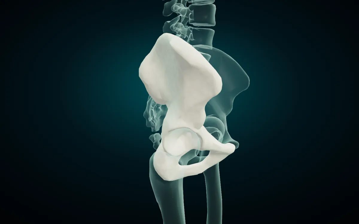
The hip bone starts life as three bones. TeachMeAnatomy puts the start of their fusion at 15 to 17 years, and OpenStax says they are one bone by the late teens. Before that, a Y-shaped plate of cartilage, the triradiate cartilage, keeps them apart inside the hip socket.
| English | Latin (TA98) | Arabic | Where it sits |
|---|---|---|---|
| Ilium | Os ilium | الحرقفة (عظم الحرقفة) | upper part, the wide flat wing you feel at your waist |
| Ischium | Os ischii | الإسك | lower back part, the bone you sit on |
| Pubis | Os pubis | عظم العانة | lower front part, meeting its partner on the midline |
| Acetabulum | Acetabulum | الحُق | the socket for the head of the femur, where all three meet |
All three bones meet in the acetabulum, the deep cup on the outer side of the hip bone. The Dartmouth-hosted Basic Human Anatomy text gives their shares: the ilium forms about the upper two fifths of the socket, the ischium about the back two fifths, and the pubis about the front fifth.
The landmarks below are the ones anatomy and physiotherapy exams ask for. Latin terms are from TA98. The Arabic follows the Arabic anatomy entry for the hip bone; for the auricular surface and the pubic tubercle it follows the atlas's own wording, which renders auricular as أذني and tubercle as حديبة elsewhere in the model.
| Part | Landmark | Latin | Arabic | Why it matters |
|---|---|---|---|---|
| Ilium | Iliac crest | Crista iliaca | عرف الورك | the upper rim you rest your hands on |
| Ilium | Anterior superior iliac spine | Spina iliaca anterior superior | الشوكة الحرقفية الأمامية العلوية | front end of the crest, a standard surface landmark |
| Ilium | Auricular surface | Facies auricularis | السطح الأذني | joins the sacrum at the sacroiliac joint |
| Ilium | Greater sciatic notch | Incisura ischiadica major | الثلمة الوركية الكبرى | the deep notch at the back of the bone |
| Ischium | Ischial tuberosity | Tuber ischiadicum | أحدوبة الإسك | takes your weight when you sit |
| Ischium | Ischial spine | Spina ischiadica | الشوكة الإسكية | separates the greater and lesser sciatic notches |
| Pubis | Pubic tubercle | Tuberculum pubicum | الحديبة العانية | small bump on the upper edge of the pubis |
| Ischium and pubis | Obturator foramen | Foramen obturatum | الثقبة السدادية | the large opening below the acetabulum |
The functions in the last column follow OpenStax, The Pelvic Girdle and Pelvis. The atlas models the adult hip bone as one part, "Hip bone", with no separate ilium, ischium or pubis to tap, and the landmarks in this table are not separate parts either. You read them off the shape. Isolate the hip bone, turn on X-ray, and turn the model to its side as in the render above: the flat wing at the top is the ilium, the cup in the middle is the acetabulum, and the ring of bone around the obturator foramen below it is the ischium behind and the pubis in front.
The pelvis as a whole
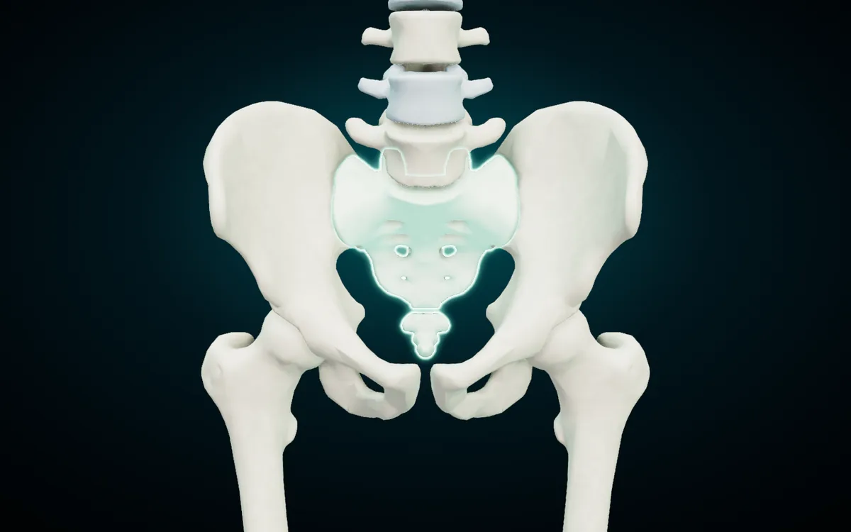
The bony pelvis is a ring of four bones: the two hip bones at the sides and front, and the sacrum and coccyx at the back. The hip bones belong to the appendicular skeleton. The sacrum and coccyx are the bottom of the vertebral column, so they belong to the axial skeleton.
| English | Latin | Arabic | What it is |
|---|---|---|---|
| Sacrum | Os sacrum | العجز | five sacral vertebrae fused into one bone |
| Coccyx | Os coccygis | العصعص | the tailbone, usually four small vertebrae fused together |
The ring is closed by joints. Each hip bone meets the sacrum at a sacroiliac joint, through its auricular surface, and the two pubic bones meet at the front at the pubic symphysis (الارتفاق العاني). The atlas's Joints system shows the pubic symphysis with its interpubic disc, and the ligaments of the sacroiliac joint. OpenStax splits the space inside into the wide greater pelvis above and the narrow lesser pelvis below. The pelvic brim, the line from the sacral promontory around to the top of the pubic symphysis, is the border between the two.
Male and female pelvis
The male and female pelvis differ in ways an examiner can ask about in one table. These differences are from OpenStax, Table 8.1:
| Feature | Female pelvis | Male pelvis |
|---|---|---|
| Bones | lighter and thinner | thicker and heavier |
| Pelvic inlet | round or oval | heart-shaped |
| Lesser pelvic cavity | shorter and wider | longer and narrower |
| Subpubic angle | greater than 80 degrees | less than 70 degrees |
| Pelvic outlet | rounded and larger | smaller |
OpenStax adds that in the female pelvis the ischial tuberosities sit farther apart and the sacrum is wider, shorter and less curved, while in the male pelvis the greater sciatic notch is narrower and deeper. The atlas has a single skeleton, so compare these features against your lab specimens or textbook figures rather than against the model.
The femur
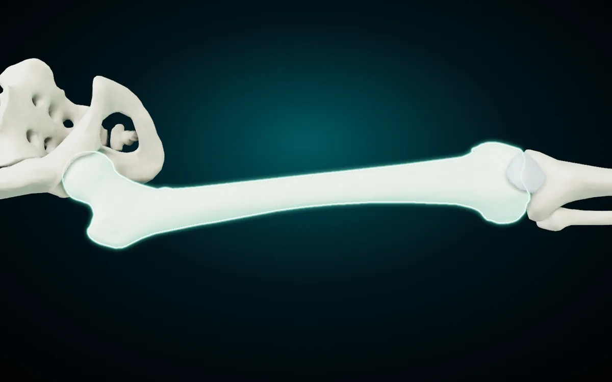
The femur (عظم الفخذ) is the thigh bone, and OpenStax calls it the longest and strongest bone of the body. Its head fits into the acetabulum to form the hip joint, and its lower end sits on the tibia at the knee. TA98 gives two Latin names for it: femur as the main term and os femoris as the official synonym. The atlas shows Os femoris.
| Landmark | Latin | Arabic | What to know |
|---|---|---|---|
| Head | Caput femoris | رأس الفخذ | fits into the acetabulum |
| Fovea of the head | Fovea capitis femoris | نقرة رأس الفخذ | pit for the ligament of the head of the femur |
| Neck | Collum femoris | عنق الفخذ | joins head to shaft; a common fracture site |
| Greater trochanter | Trochanter major | المدور الكبير | large lateral bump for hip muscles |
| Lesser trochanter | Trochanter minor | المدور الصغير | smaller medial bump below the neck |
| Linea aspera | Linea aspera | الخط الخشن | ridge down the back of the shaft for muscle attachments |
| Medial and lateral condyles | Condylus medialis / lateralis | اللقمة الإنسية / الوحشية | rounded ends that sit on the tibia |
| Medial and lateral epicondyles | Epicondylus medialis / lateralis | اللقيمة الإنسية / الوحشية | bumps above the condyles for knee ligaments |
| Patellar surface | Facies patellaris | السطح الرضفي | groove at the front where the patella glides |
| Intercondylar fossa | Fossa intercondylaris | الحفرة بين اللقمتين | deep notch between the condyles at the back |
Descriptions follow OpenStax; Latin terms are from TA98 and Arabic from the Arabic anatomy entry for the femur. As with the hip bone, the atlas models the femur as one part. To find the landmarks, isolate the femur and turn it: the greater trochanter is the bump on the outer side below the hip, the lesser trochanter points inward and back, and the linea aspera shows as a raised line when you look at the back of the shaft.
The patella
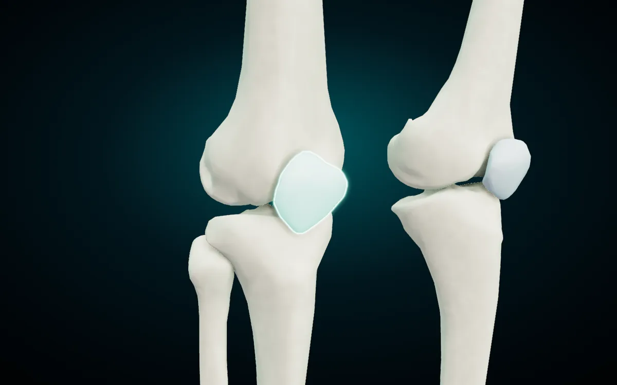
The patella (الرضفة), the kneecap, is the largest sesamoid bone in the body. It grows inside the tendon of the quadriceps femoris and glides in the patellar surface of the femur. OpenStax points out a detail students often miss: the patella articulates only with the femur, not with the tibia. Below the patella the same tendon continues to the tibial tuberosity.
The knee joint itself lives in the atlas's Joints system. Turn it on and the medial and lateral menisci, the anterior and posterior cruciate ligaments, and the tibial and fibular collateral ligaments each get their own card in all three languages.
The tibia and the fibula
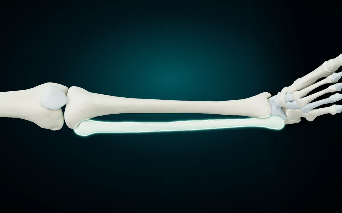
The leg below the knee has two bones. The tibia (الظنبوب), the shin bone, is on the medial side, carries the body's weight to the foot, and is the second longest bone in the body after the femur. The fibula (الشظية) is the slim bone on the lateral side. OpenStax states that the fibula does not bear weight, and it serves mainly for muscle attachments. A sheet of tissue, the interosseous membrane, joins the two bones along their length.
| Bone | Landmark | Latin | Arabic | What to know |
|---|---|---|---|---|
| Tibia | Medial and lateral condyles | Condylus medialis / lateralis | اللقمة الإنسية / الوحشية | flat tops that take the femoral condyles |
| Tibia | Intercondylar eminence | Eminentia intercondylaris | البروز بين اللقمتين | ridge between the condyles where the cruciate ligaments attach |
| Tibia | Tibial tuberosity | Tuberositas tibiae | الأحدوبة الظنبوبية | bump below the knee where the patellar tendon attaches |
| Tibia | Anterior border | Margo anterior | الحافة الأمامية | the sharp shin you can feel under the skin |
| Tibia | Medial malleolus | Malleolus medialis | الكعب الإنسي | the inner ankle bump |
| Fibula | Head | Caput fibulae | رأس الشظية | knob below the lateral tibial condyle |
| Fibula | Lateral malleolus | Malleolus lateralis | الكعب الوحشي | the outer ankle bump |
Facts are from OpenStax; Latin from TA98; Arabic from the Arabic anatomy entry for the tibia and from the atlas itself, which uses رأس الشظية in the names of the fibular head ligaments and الكعب الإنسي and الكعب الوحشي for the two ankle bumps. The two malleoli hold the talus between them like a clamp, and that is the ankle joint. In the atlas, "Medial malleolus" and "Lateral malleolus" are skin areas in the Regions system, not parts of the bones, so a search for malleolus lands on the surface of the ankle. To see the bony malleoli, look at the lower ends of the tibia and fibula on the skeleton.
Bones of the ankle and foot
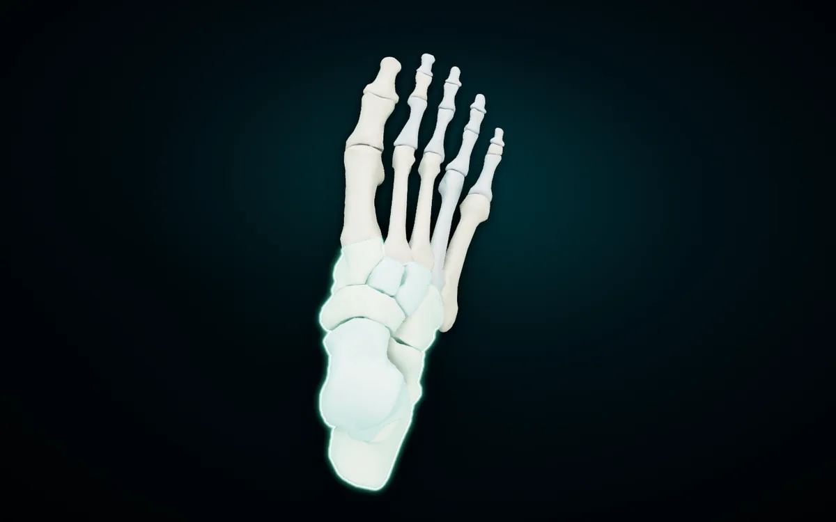
Each foot has 26 bones: 7 tarsals, 5 metatarsals and 14 phalanges. TeachMeAnatomy sorts the tarsals into three rows, and that grouping is the easiest way to hold them in your head:
| Row | English | Latin | Arabic |
|---|---|---|---|
| Proximal | Talus | Talus (atlas: Os tali) | الكاحل |
| Proximal | Calcaneus | Calcaneus | العقب |
| Intermediate | Navicular bone | Os naviculare | العظم الزورقي |
| Distal | Medial cuneiform bone | Os cuneiforme mediale | العظم الإسفيني الإنسي |
| Distal | Intermediate cuneiform bone | Os cuneiforme intermedium | العظم الإسفيني المتوسط |
| Distal | Lateral cuneiform bone | Os cuneiforme laterale | العظم الإسفيني الوحشي |
| Distal | Cuboid bone | Os cuboideum | العظم النردي |
- Talus. The top bone of the foot. It sits between the two malleoli to form the ankle joint, rests on the calcaneus below and meets the navicular in front. TeachMeAnatomy notes that no muscles attach to it.
- Calcaneus. The heel bone and the largest tarsal. The Achilles tendon attaches to its back.
- Navicular. The boat-shaped bone on the inner side of the foot, between the talus and the three cuneiforms.
- Cuneiforms. Three wedge-shaped bones in a row, medial, intermediate and lateral, each in front of the navicular and behind a metatarsal.
- Cuboid. The outermost bone of the distal row, in front of the calcaneus and behind the fourth and fifth metatarsals.
The same rows give the three regions of the foot. The talus and calcaneus make the hindfoot, the navicular, cuboid and cuneiforms the midfoot, and the metatarsals and phalanges the forefoot.
Metatarsals and phalanges
| English | Latin (atlas) | Arabic |
|---|---|---|
| First metatarsal bone | Os primum metatarsi | عظم مشط القدم الأول |
| Second metatarsal bone | Os secundum metatarsi | عظم مشط القدم الثاني |
| Third metatarsal bone | Os tertium metatarsi | عظم مشط القدم الثالث |
| Fourth metatarsal bone | Os quartum metatarsi | عظم مشط القدم الرابع |
| Fifth metatarsal bone | Os quintum metatarsi | عظم مشط القدم الخامس |
| Proximal phalanx | Phalanx proximalis | السلامية الدانية |
| Middle phalanx | Phalanx media | السلامية الوسطى |
| Distal phalanx | Phalanx distalis | السلامية البعيدة |
The metatarsals are numbered 1 to 5 from the big-toe side. OpenStax notes that the first is shorter and thicker than the rest and the second is the longest. Their heads form the ball of the foot.
The toes follow the pattern of the fingers. The big toe, the hallux, has two phalanges, proximal and distal. The other four toes have three each, which gives 2 + 4 × 3 = 14. The atlas names every phalanx by row and toe in all three languages, for example "Distal phalanx of fifth finger of foot", Phalanx distalis digiti quinti pedis, السلامية البعيدة لإصبع القدم الخامس.
The atlas also has a part called "Sesamoid bones of foot" (العظام السمسمانية للقدم). These are the two small bones under the head of the first metatarsal, which the Basic Human Anatomy text says are almost always present. The usual count of 206 leaves them out, though it includes the patella.
The arches of the foot
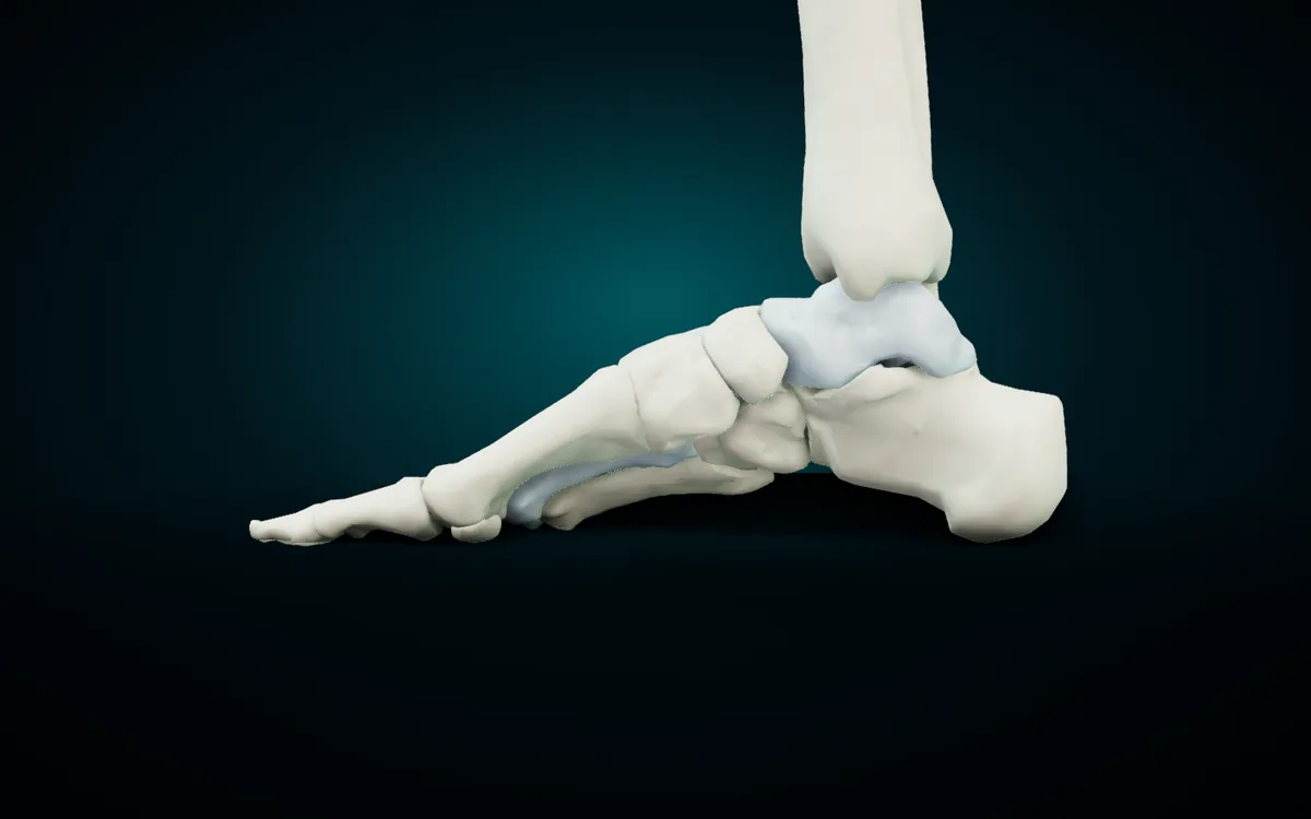
From the inner side, the bones of the foot form a curve. OpenStax describes two longitudinal arches and one transverse arch:
- Medial longitudinal arch. The higher of the two, along the inner side of the foot. The calcaneus holds it at the back, the metatarsal heads at the front, and the talus sits at its top. It is the curve in the render above.
- Lateral longitudinal arch. Along the outer side, and much flatter.
- Transverse arch. Across the foot, formed by the cuneiforms and the bases of the metatarsals.
The arches flatten a little under load and spring back, which absorbs shock and stores energy in the ligaments. When the ligaments stretch over time and the arches fall, mostly on the medial side, the result is a flat foot, pes planus. In the atlas, the Regions system marks the medial and lateral parts of the longitudinal arch and the proximal and distal transverse arches as areas on the skin of the foot, named in all three languages.
Latin and Arabic traps in the leg and foot
Talus or os tali. TA98 gives the talus a one-word Latin name, talus. The atlas card reads Os tali, "bone of the talus". If your exam asks for the TA term, write talus.
Femur or os femoris. Both are correct. TA98 lists femur as the preferred term and os femoris as an official synonym.
Numbered metatarsals. TA98 lists the metatarsals as one group, Ossa metatarsi [I–V], without separate Latin entries for each bone. The atlas uses Os primum metatarsi for the first, as the IMAIOS e-Anatomy entry does, and other references write Os metatarsale I. Each version names the same bone.
"Finger of foot". The atlas's English names for the toe phalanges say "finger of foot" where most textbooks say "toe": "Proximal phalanx of first finger of foot" is the proximal phalanx of the big toe. The Latin digiti primi pedis means the same thing. Search for phalanx or for the Arabic إصبع القدم to list them.
الكاحل is not the whole ankle. In the atlas, الكاحل is the talus. In everyday Arabic the same word means the ankle region, so read it in context.
Two boat-shaped bones. Arabic calls both the navicular of the foot and the scaphoid of the wrist العظم الزورقي. The atlas adds (الرسغ), "wrist", to the scaphoid to keep them apart.
Let the meaning carry the name. Several Arabic names translate the Latin: cuneus means wedge and إسفين means wedge, calx means heel and العقب means heel, and the cuboid is cube-shaped while نرد is a die. Learn one and the other follows.
How to memorise the leg and foot bones
Count first. One hip bone, then one bone in the thigh, one in front of the knee, two in the leg, and 26 in the foot. Write 1 + 1 + 1 + 2 + 26 = 31 at the top of your page and check your list adds up.
Use one mnemonic for the tarsals. "Tiger Cubs Need MILC" gives Talus, Calcaneus, Navicular, Medial, Intermediate and Lateral cuneiforms, Cuboid. It follows the rows in the table above, from the ankle forward and from the inner side out.
Tie each bone to something you can feel. The iliac crest at your waist, the ischial tuberosities under you when you sit, the greater trochanter on the side of your hip, the patella, the sharp anterior border of the tibia, the two malleoli at the ankle, and the calcaneus at your heel are all under the skin. Touch each one while you say its three names.
Check yourself on the model. Read a name from the tables, find the bone on the 3D foot, and let the card confirm it.
Find each bone in the 3D atlas
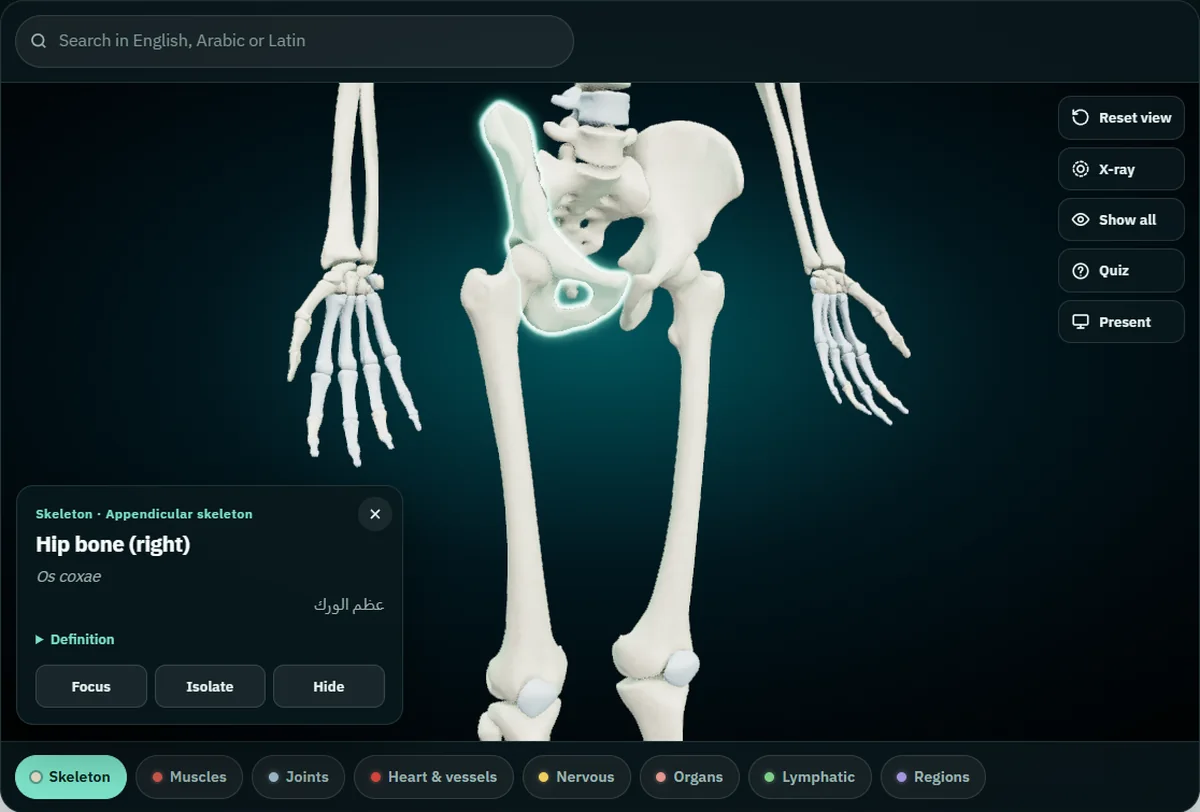
The free 3D skeleton runs in the browser on a laptop, tablet or phone, with no account. The skeleton loads first. A route from the hip to the toes:
- Fly to the pelvis. Double-tap a hip bone and the camera moves in close. Drag to turn the model, pinch or scroll to zoom.
- Tap each bone. A card opens with the English name and side, the Latin term and the Arabic name, plus a short definition. Paired bones carry their side, "Hip bone (right)" in English and "(يمين)" in the Arabic version of the atlas. The card also gives the group: the hip bone, femur and foot bones read "Appendicular skeleton".
- Isolate the hip bone, then X-ray. Press Isolate on its card, then X-ray. The hip bone stays solid and the rest of the skeleton turns to glass, so you see the acetabulum around the head of the femur. Turn the model to its side to read the ilium, ischium and pubis.
- Search in any of the three languages. Type cuboid, os cuboideum or النردي and the model flies to the bone and opens its card. Search is the fastest way to reach the three cuneiforms, which sit close together.
- Hide the leg bones to see the foot. Tap the tibia and press Hide, then the fibula. Turn the foot so the toes point away from you and look down on the tarsals from above, as in the foot render. Show all brings everything back.
- Turn on Joints for the knee and hip. The Joints chip adds the acetabular labrum, the pubic symphysis, the menisci and the cruciate and collateral ligaments, each with its own three-language card.
- Quiz yourself. Quiz asks ten "Where is the…?" questions and you answer by tapping the model. It draws from every part still on screen, so with the full skeleton showing, the leg is only part of the pool. For a leg-and-foot drill, turn the leg toward you, read a name from the tables above, tap where you think it is, and let the card tell you if you were right.
Present mode fills the screen and names each tapped bone in large type in all three languages, which suits a study group around one screen. The guide to teaching anatomy with a projector covers that setup. When the bones are solid, the muscles of the human body in English, Latin and Arabic use the same three-column format, and the hip, thigh and leg muscles attach to the landmarks in this guide.
Frequently asked questions
How many bones are in the leg and foot?
30 in each lower limb: femur, patella, tibia, fibula, 7 tarsals, 5 metatarsals and 14 phalanges. Count the hip bone as well and it is 31 per side. The foot alone has 26.
What are the three parts of the hip bone?
The ilium above, the ischium at the back and below, and the pubis at the front. They are separate bones in childhood, start to fuse in the mid to late teens, and meet in the acetabulum, the socket for the femur.
Which is the longest bone in the body?
The femur. It is also the strongest. The tibia is the second longest.
Does the fibula carry body weight?
No. The tibia carries the weight from the knee to the ankle. The fibula is mainly a site for muscle attachments, and its lower end forms the lateral malleolus of the ankle.
Which is the largest tarsal bone?
The calcaneus, the heel bone. The talus sits on top of it and forms the ankle joint with the tibia and fibula.
Is the patella counted among the 206 bones?
Yes. It is the largest sesamoid bone in the body and part of the usual count. The two small sesamoid bones under the big toe are not counted.
Why does the atlas say Os tali when my book says talus?
TA98 uses the single word talus, and that is the term to write in an exam that follows Terminologia Anatomica. The atlas shows Os tali. Both name the same bone.
Start with the hip bone in 3D
Open the 3D pelvis and leg bones in your browser, isolate the hip bone and find the acetabulum. Then work down: femur, patella, tibia and fibula, and the seven tarsals by row. Once you can name all 31 without the card, hide a few and run the quiz. If you study on your phone, Anatomy 3D for Android carries the same models and names, works offline and adds cut planes on three axes; the Anatomy 3D app page has the details.
3D renders: NasrTech, from Z-Anatomy models (CC BY-SA 4.0).