

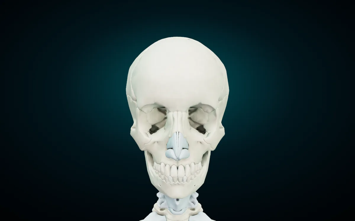
The adult skull has 22 bones: 8 cranial bones that enclose the brain and 14 facial bones that build the face. This guide names every one of them in English, Latin and Arabic, says where each sits and what it touches, and then covers the sutures that lock them together, the fontanelles of a newborn, and a short way to memorise the list. The names in the tables are the ones the free 3D Anatomy Atlas shows when you tap a bone, and every picture is a render of that same model.
For the rest of the skeleton (spine, ribs, limbs, hands and feet) see the overview of the bones of the human body in English, Latin and Arabic. This page covers the head in more depth.
The 22 skull bones at a glance
| Group | Bone | Paired? | Count |
|---|---|---|---|
| Cranial | Frontal bone | single | 1 |
| Cranial | Parietal bone | paired | 2 |
| Cranial | Occipital bone | single | 1 |
| Cranial | Temporal bone | paired | 2 |
| Cranial | Sphenoid bone | single | 1 |
| Cranial | Ethmoid bone | single | 1 |
| Facial | Nasal bone | paired | 2 |
| Facial | Maxilla | paired | 2 |
| Facial | Zygomatic bone | paired | 2 |
| Facial | Lacrimal bone | paired | 2 |
| Facial | Palatine bone | paired | 2 |
| Facial | Inferior nasal concha | paired | 2 |
| Facial | Vomer | single | 1 |
| Facial | Mandible | single | 1 |
| Total | 22 |
That is the split used in standard teaching texts such as OpenStax Anatomy and Physiology: eight bones in the brain case and fourteen in the face. Six more bones sit inside the skull, the three ear ossicles on each side, and textbooks count them separately. The hyoid hangs in the neck below the mandible and is not a skull bone at all. Both are covered in their own section below.
One pattern makes the table easy to hold in your head. In the cranium, only two bones come in pairs: the parietal and the temporal. In the face it is the reverse, and only two bones are single: the vomer and the mandible.
The 8 cranial bones
| English | Latin | Arabic | Paired? |
|---|---|---|---|
| Frontal bone | Os frontale | العظم الجبهي | single |
| Parietal bone | Os parietale | العظم الجداري | paired |
| Occipital bone | Os occipitale | العظم القذالي | single |
| Temporal bone | Os temporale | العظم الصدغي | paired |
| Sphenoid bone | Os sphenoideum | العظم الوتدي | single |
| Ethmoid bone | Os ethmoideum | العظم الغربالي | single |
What each one does and borders:
- Frontal bone. The forehead and the roofs of both orbits. It holds the frontal sinus above the eyebrows and meets the two parietal bones at the coronal suture.
- Parietal bones. The roof and upper sides of the cranium. The right and left parietal bones meet each other on the midline at the sagittal suture.
- Occipital bone. The back and much of the base of the skull. The spinal cord passes through its large opening, the foramen magnum, and its two occipital condyles rest on the first cervical vertebra (C1).
- Temporal bones. The lower sides and part of the base, around each ear. The petrous part holds the middle and inner ear. The mastoid process anchors muscles behind the ear, the styloid process carries a ligament of the hyoid, and the mandibular fossa takes the head of the mandible to form the jaw joint.
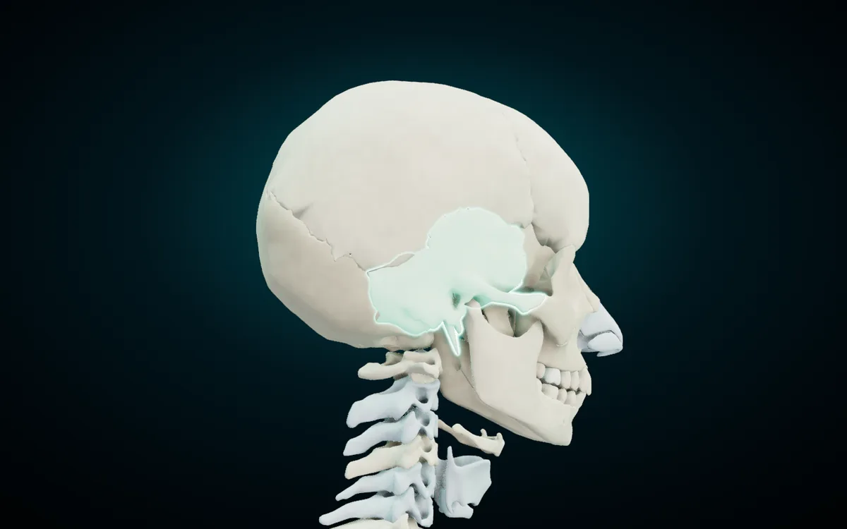
- Sphenoid bone. A single bone that runs across the middle of the cranial floor. OpenStax calls it a "keystone" bone because it joins with almost every other bone of the skull. A saddle on its upper surface, the sella turcica, holds the pituitary gland, and its body contains the sphenoid sinus.
- Ethmoid bone. A light, box-shaped bone between the two orbits. Its cribriform plate forms part of the floor of the anterior cranial cavity and lets the olfactory nerve fibres through (the first of the 12 cranial nerves), its perpendicular plate forms the upper part of the nasal septum, and its sides carry the superior and middle nasal conchae and the ethmoid air cells.
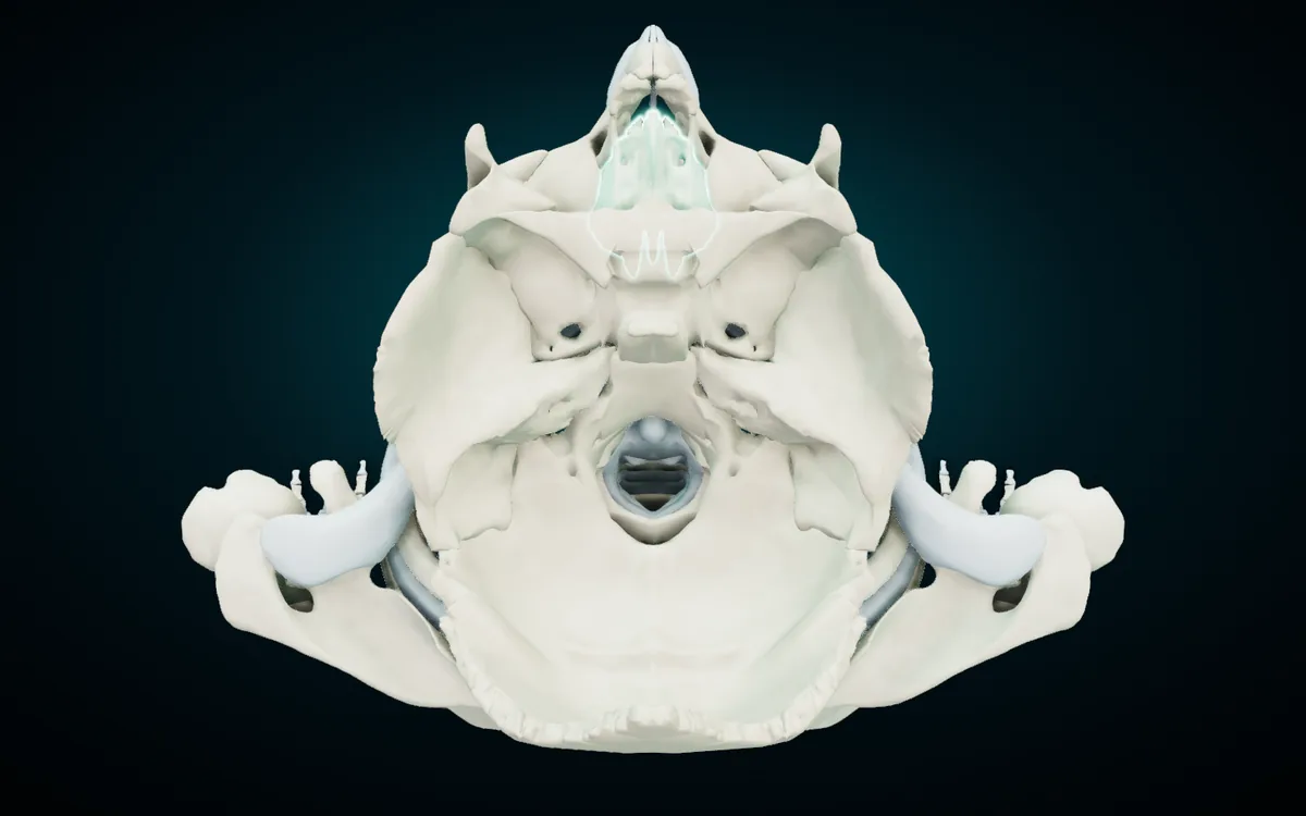
The sphenoid and the ethmoid are the two bones students struggle to picture, because from outside you see only small patches of them. The render above is what you get in the atlas after you hide the frontal bone, both parietal bones and the frontal sinus (a separate part on this model) and look down into the skull. The front of the skull is at the top of the picture. The ethmoid (highlighted) sits at the front between the orbits, the sphenoid spreads across the middle, the petrous parts of the temporal bones angle in from each side, and the occipital bone wraps around the foramen magnum at the back.
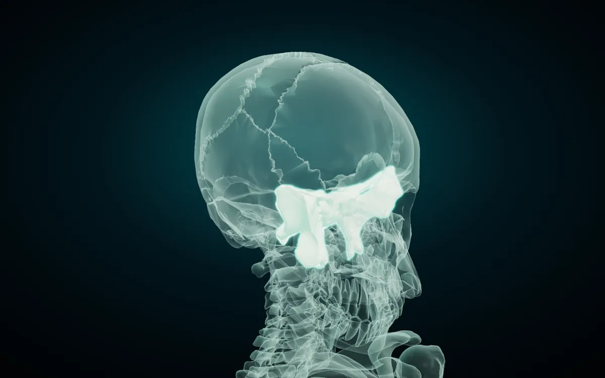
Isolate the sphenoid and turn on X-ray, and the rest of the skull becomes glass while the sphenoid stays solid. Now you can see why it is described as wing-shaped: a central body, a greater and lesser wing on each side, and two pterygoid processes hanging down behind the maxillae.
The 14 facial bones
| English | Latin | Arabic | Paired? |
|---|---|---|---|
| Nasal bone | Os nasale | العظم الأنفي | paired |
| Maxilla | Maxilla | الفك العلوي | paired |
| Zygomatic bone | Os zygomaticum | العظم الوجني | paired |
| Lacrimal bone | Os lacrimale | العظم الدمعي | paired |
| Palatine bone | Os palatinum | العظم الحنكي | paired |
| Inferior nasal concha | Concha nasalis inferior | المحارة الأنفية السفلية | paired |
| Vomer | Vomer | الميكعة | single |
| Mandible | Mandibula | الفك السفلي | single |
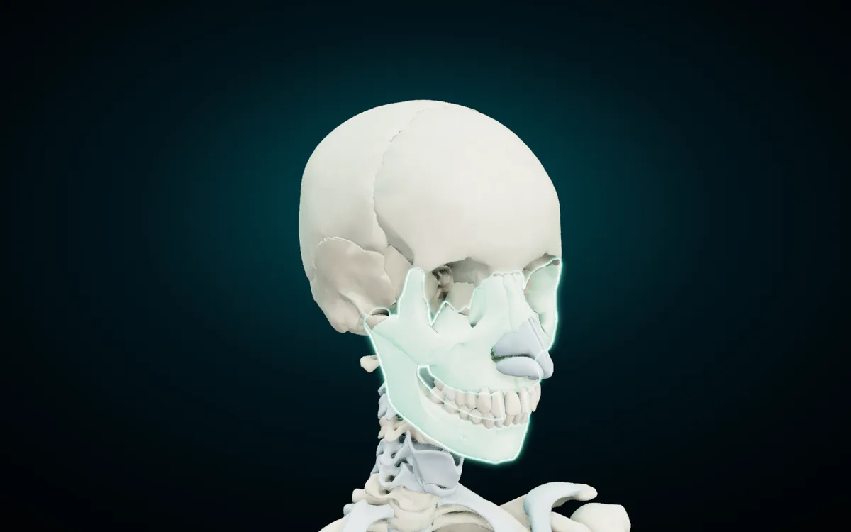
- Nasal bones. Two small oblong bones side by side that form the bony bridge of the nose. The rest of the external nose is cartilage, and the atlas models those cartilages too.
- Maxillae. The two maxillae form the upper jaw and carry the upper teeth. They make up most of the hard palate and the floor of each orbit, and each holds a maxillary sinus, the largest of the paranasal sinuses.
- Zygomatic bones. The cheekbones. Each forms part of the outer wall and floor of the orbit, and its temporal process joins the zygomatic process of the temporal bone to make the zygomatic arch.
- Lacrimal bones. Small, thin bones in the front of the inner wall of each orbit, next to the tear duct. The atlas definition puts one at roughly the size of the little fingernail, and it is usually described as the smallest and most fragile bone of the face.
- Palatine bones. L-shaped bones at the back of the nasal cavity. Their horizontal plates form the back part of the hard palate, behind the maxillae.
- Inferior nasal conchae. Curled plates on the outer wall of each nasal cavity. They are independent bones. The superior and middle conchae above them are not, because they belong to the ethmoid.
- Vomer. A thin, single bone that forms the lower and back part of the nasal septum, below the perpendicular plate of the ethmoid.
- Mandible. The lower jaw, carrying the lower teeth. Of the 22 skull bones it is the only one that moves at a joint: the temporomandibular joint, where its condyle sits in the mandibular fossa of the temporal bone.
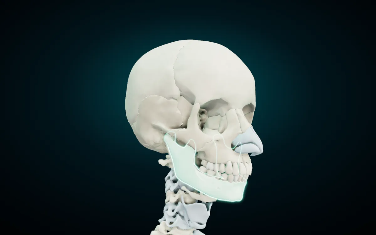
Dental students get one extra on this model: each tooth is its own tappable part, named in all three languages, so the teeth of the maxilla and the mandible can be studied on the same skull as the bones that hold them.
Ear ossicles and the hyoid
| English | Latin | Arabic | Where |
|---|---|---|---|
| Malleus | Malleus | المطرقة | middle ear, attached to the eardrum |
| Incus | Incus | السندان | middle ear, between malleus and stapes |
| Stapes | Stapes | الركاب | middle ear, at the oval window of the inner ear |
| Hyoid bone | Os hyoideum | العظم اللامي | neck, below the mandible |
The three ossicles sit in the middle ear inside each temporal bone and pass sound vibrations from the eardrum to the inner ear. The stapes is the smallest bone in the body. With six ossicles on top of the 22, some books give 28 bones for the head. Add the hyoid and you reach 29, but the hyoid does not belong to the skull: it touches no other bone and is held in place by muscles and ligaments. In the atlas the ossicles carry the group "Auditory ossicles" on their card, and the stylohyoid ligament that ties the hyoid to the temporal bone is in the Joints system.
Sutures: where the cranial bones meet
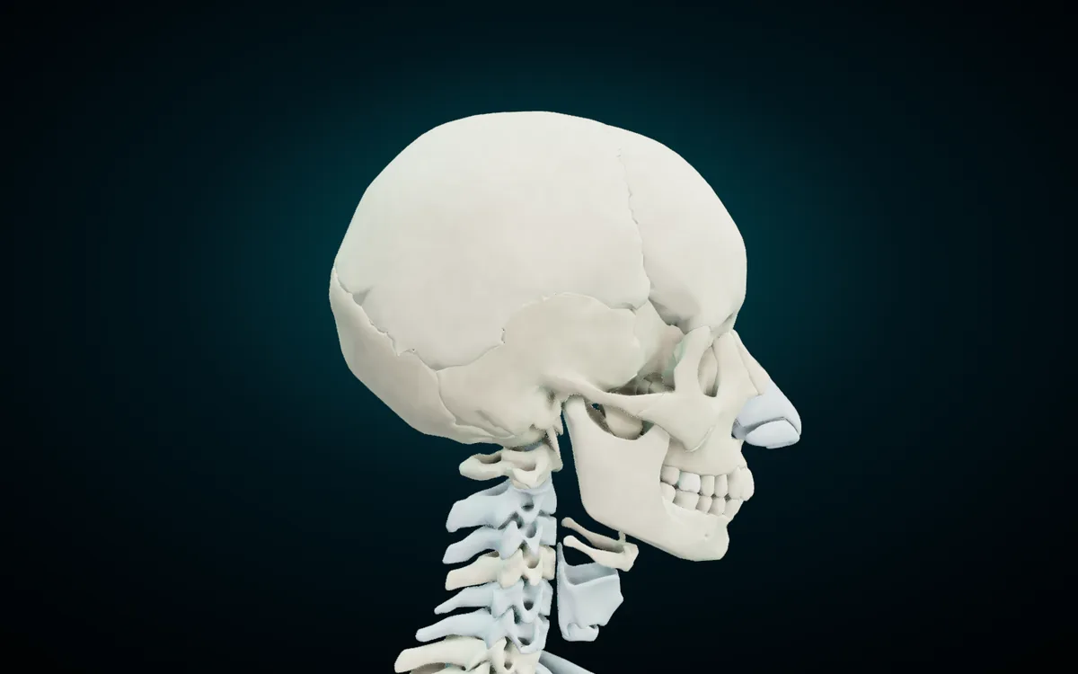
Cranial bones are joined by sutures, fibrous joints that hardly move in an adult. Four of them come up in every exam.
| English | Latin | Arabic | Joins |
|---|---|---|---|
| Coronal suture | Sutura coronalis | الدرز الإكليلي | frontal bone and both parietal bones |
| Sagittal suture | Sutura sagittalis | الدرز السهمي | right and left parietal bones |
| Lambdoid suture | Sutura lambdoidea | الدرز اللامي | occipital bone and both parietal bones |
| Squamous suture | Sutura squamosa | الدرز الحرشفي | temporal bone and parietal bone |
Sources for the joints: OpenStax, The Skull; the Arabic terms follow the Arabic anatomy list of named cranial sutures.html). The atlas does not model sutures as separate parts, so you won't find them by search, but you can see the suture lines on the bones themselves. Turn on X-ray and the zigzag edges of the frontal, parietal and occipital bones stand out, as in the sphenoid render above.
One meeting point on the side of the head has its own name: the pterion, where the frontal, parietal, temporal and sphenoid bones meet. OpenStax calls it the weakest part of the skull. An artery runs just inside it, and a blow there can fracture the thin bone, tear the artery and cause a bleed that presses on the brain. To find it on the model, turn the skull to its side and tap around the temple, just above the zygomatic arch, where the edges of those four bones come together.
Fontanelles: the soft spots of a newborn skull
At birth the cranial bones have not met yet. The gaps between them are covered by membrane, and the larger gaps are called fontanelles (اليافوخ in Arabic). They let the skull mould during birth and let it grow with the brain.
| Fontanelle | Latin | Where | Usually closes |
|---|---|---|---|
| Posterior | Fonticulus posterior | where the sagittal and lambdoid sutures meet | about 2 to 3 months after birth |
| Sphenoidal (anterolateral) | Fonticulus sphenoidalis | on the side, at the pterion | around 6 months |
| Mastoid (posterolateral) | Fonticulus mastoideus | behind the ear, where parietal, occipital and temporal meet | 6 to 18 months |
| Anterior | Fonticulus anterior | where the coronal and sagittal sutures meet | last to close, between 1 and 3 years |
Ages are from the CARTA anthropogeny summary of fontanelle closure, which also notes wide variation from child to child. The anterior fontanelle is the diamond-shaped soft spot on top of a baby's head. Its adult landmark is the bregma, and the posterior fontanelle becomes the lambda. The atlas shows an adult skull, so the fontanelles are closed on the model. Use their landmarks instead: find where the coronal and sagittal suture lines cross on top of the head, and you are on the site of the anterior fontanelle.
Four skull bones with sinuses
The paranasal sinuses are air spaces inside four of the skull bones: the frontal bone, the two maxillae, the sphenoid and the ethmoid. The atlas gives two of them their own entries, "Sinus of frontal bone" (الجيب الجبهي) and "Sinus of sphenoid bone" (الجيب الوتدي). Hide the frontal bone and the frontal sinus stays behind as its own shape above the orbits, which shows how far it reaches.
Two Latin spellings you will meet
Two cranial bones appear with two Latin endings, depending on the book.
- The atlas shows Os sphenoideum and Os ethmoideum. These are the terms in Terminologia Anatomica 2nd edition (TA2), the current international list.
- The 1998 edition (TA98) and many textbooks print Os sphenoidale and Os ethmoidale. The TA98 entry for the sphenoid uses that form, and TA2 keeps it as a synonym.
Both spellings name the same bone. If your department follows one edition, use its spelling.
How to memorise the skull bones
Use the mnemonics, then check the count. Cranial bones: PEST OF 6, for Parietal, Ethmoid, Sphenoid, Temporal, Occipital, Frontal. Six names, eight bones, because parietal and temporal come in pairs. Facial bones: "Virgil Can Not Make My Pet Zebra Laugh", for Vomer, Conchae (inferior nasal), Nasal, Maxilla, Mandible, Palatine, Zygomatic, Lacrimal. Eight names, fourteen bones, because all but the vomer and mandible come in pairs.
Let the Arabic name explain the Latin one. Most Arabic skull names translate the meaning of the Latin or Greek word, so one language reminds you of the other:
| Latin or Greek root | Meaning | Arabic name | Arabic word means |
|---|---|---|---|
| paries (parietal) | wall | الجداري | جدار, wall |
| sphen (sphenoid) | wedge | الوتدي | وتد, peg or wedge |
| ethmos (ethmoid) | sieve | الغربالي | غربال, sieve |
| lacrima (lacrimal) | tear | الدمعي | دمع, tears |
| concha | shell | المحارة | محارة, shell |
Once you know that the ethmoid is "the sieve bone", its cribriform plate full of holes makes sense in both languages. The same goes for the vomer, from the Latin for ploughshare, which is roughly its shape in the nasal septum.
Learn on the model, not the list. Say each bone's name out loud as you tap it, then close the card and point to it again from a different angle.
Find each bone in the 3D atlas
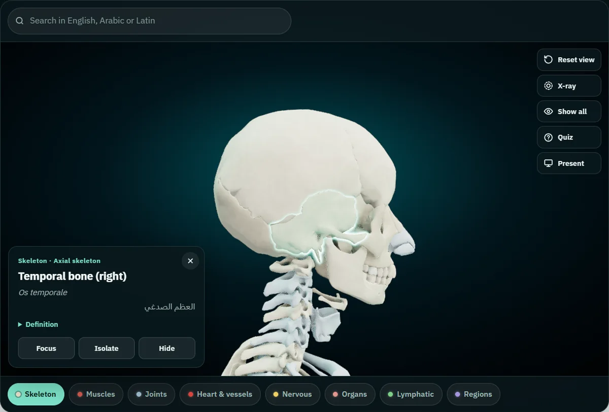
The free 3D skull and skeleton runs in the browser on a laptop, tablet or phone, with no account. The skeleton loads first. Here is a route through the 22 bones:
- Fly to the skull. Double-tap the head and the camera moves in close. Drag to turn the model and pinch or scroll to zoom.
- Tap each bone. A card opens with the English name and side, the Latin term and the Arabic name, plus a short definition. Paired bones carry their side: "Temporal bone (right)" in English, "(يمين)" in the Arabic version of the atlas. On a computer, hovering over a bone shows its name before you click.
- Search in any of the three languages. Type sphenoid, os sphenoideum or الوتدي, pick the result, and the model flies to the bone and opens its card. This is the quickest way to reach the lacrimal, palatine and inferior nasal concha, which hide behind other bones.
- Hide what is in the way. Tap the frontal bone and press Hide, then do the same for both parietal bones. You are now looking into the cranial cavity at the floor shown earlier in this guide. Show all brings everything back.
- Isolate, then X-ray. Select the sphenoid or the ethmoid, press Isolate, then press X-ray. The chosen bone stays solid and the rest of the skull turns to glass, so you see its shape and where it sits at the same time.
- Quiz yourself. Quiz asks ten "Where is the…?" questions and you answer by tapping the model. It draws from every part still on screen, so with the full skeleton showing, the skull is only part of the pool. For a skull-only drill, turn the head toward you, read a name from the tables above, tap where you think it is, and let the card tell you if you were right.
Present mode fills the screen and names each tapped bone in large type in all three languages, which suits a study group around one screen or a lecturer at the front of the room. The guide to teaching anatomy with a projector covers that setup. When the bones are solid, the muscles of the human body in English, Latin and Arabic use the same three-column format, with a section on the head and neck.
Frequently asked questions
How many bones are in the human skull?
22: eight cranial bones and fourteen facial bones. Some books count the six middle-ear ossicles with the skull, which gives 28. The hyoid is not part of the skull.
Which skull bone can move?
Only the mandible. It moves at the temporomandibular joint, on each side, in front of the ear. All the other skull bones are held together by sutures and other fibrous joints.
Which is the keystone bone of the skull?
The sphenoid. It sits across the middle of the cranial floor and joins with almost every other bone of the skull.
Are the superior and middle nasal conchae separate bones?
No. They are parts of the ethmoid bone. Only the inferior nasal concha is a separate bone, which is why it appears in the list of 14 facial bones.
Is the hyoid a skull bone?
No. It sits in the neck between the mandible and the larynx and does not touch any other bone. It is counted with the axial skeleton, not with the skull.
Why does the atlas say Os sphenoideum when my book says Os sphenoidale?
The atlas follows Terminologia Anatomica 2nd edition (TA2), which uses Os sphenoideum and Os ethmoideum. The older TA98 and many textbooks use Os sphenoidale and Os ethmoidale. Both are correct names for the same bone.
Start with the skull in 3D
Open the 3D skull in your browser, search for the sphenoid, and work outward from it: the ethmoid in front, the temporal bones at the sides, the occipital behind, then the face. Once you can name all 22 without the card, hide a few and test yourself again. If you study on your phone, Anatomy 3D for Android carries the same models and names, works offline and adds cut planes on three axes; the Anatomy 3D app page has the details.
Next in this series: the bones of the pelvis and leg and the 12 cranial nerves, named the same way.
3D renders: NasrTech, from Z-Anatomy models (CC BY-SA 4.0).